1. What were the three aspects of the assignments I've submitted that I am most proud of?
I would have to say that the 3 things I am most proud of are the cell model, the ethical issues essay, and the microscope lab.
2. What two aspects of my submitted assignments do I believe could have used some improvement?
Two aspects of my assignments that could have used some improvement were completing everything on time and the dragon lab.
3. What do I believe my overall grade should be for this unit?
I feel that my overall grade for unit 1 should be around 80%-85%.
4. How could I perform better in the next unit?
I can perform better in the next unit by giving my self more time to complete the assignments.
Saturday, February 23, 2008
Do You Want To Be Cloned?
Is cloning a good thing for society? Cloning is the process of duplicating biological material. There are three types of cloning technologies; recombinant DNA technology, reproductive cloning, and therapeutic cloning. Recombinant DNA technology is when a DNA fragment of interest is transferred from one organism to a self replicating genetic element like bacteria. Reproductive cloning is when an animal is made that has the same nuclear DNA as another animal. Therapeutic cloning is the production of human embryos for use in research. There are many arguments that are for and against the science of cloning.
There are many reasons why cloning can be a good thing for society. Gene therapy is used to treat certain genetic conditions. Food crops are genetically engineered to improve taste and nutritional value, and to resist certain types of diseases. Reproductive cloning is used to repopulate endangered animals or animals that are difficult to breed. Therapeutic cloning may be used to produce whole organs from single cells in humans, and to produce healthy cells to replace damaged ones in degenerative diseases.
There are also many reasons why cloning may be a bad thing for society. Reproductive cloning is expensive and very inefficient. Cloned animals have more compromised immune function and higher rate of infection, tumor growth, and other disorders. Many of the cloned animals have not lived long enough to create good data about how clones age. Many feel that human cloning is unethical because of the lack of understanding about reproductive cloning, we are not sure how cloning may impact mental development. The amount of unknown variables makes attempting to clone humans potentially dangerous and therefore ethically irresponsible.
There are both good and bad things about cloning. Cloning can be used to treat genetic conditions, repopulate certain species of animals, and may be used to produce human organs for transplant. These reasons make cloning a wonderful thing for society. There are also many reasons why cloning is not good for society. Cloning is expensive, inefficient, and clones have shown more developmental problems than normal. These reasons cause people to feel that cloning is unethical.
I feel that if there cloned animals that live longer allowing more studies and observations to be made cloning will do some amazing things in the future. If there is not success with cloning in the near future I feel that the unknown will scare society too much and cloning will be outlawed.
Monday, February 18, 2008
Unit 1 Lab Project: Build a Cell
Cell Materials

These are the different items I used to make the cell.
The Silver Bowl is the Cell Membrane
The Stuffing is the Cytoplasm
Purple Ball is the Nucleus
The Blue Ribbon is the Rough Endoplasmic Reticulum
Blue Straight Pins are the Ribosomes
The Yellow Ribbon is the Smooth Endoplasmic Reticulum
The Crackers are the Golgi Apparatus
Clear Pink Beads are the Lysosomes
Clear Yellow Vitamis E pills are the Vesicles
Yellow Vitamins are the Mitochondria
The Thread is the Chromatin
Yellow ball is the Nucleolus
Functional Cell


DNA strand is a double helix, when straightnened out it looks like a ladder. the sides of the ladder are made of phosphate-sugar backbones. The rungs of the ladder are alternating pairs of bases, thymine bonded with adenine & guanine bonded with cytosine.

The Hydrogen bonds holding the base pairs together "unzips", mRNA nucleotides are joining with RNA polymerase creating a new strand of DNA.
Mitosis

Prophase

Chromosomes are visible in the nucleus.
The nucleus fragments releasing the chromosomes.
Metaphase

Centromeres of duplicated chromosomes are aligned at the center of the cell.
Spindle Fibers attatched to the sister chromatids come from the asters on each end of the cell.
Anaphase

Sister chromatids part and become daughter chromosomes that move towards the spindle poles(aster).
Telophase

Daughter cells are forming as nuclear envelopes and nucleoli reappear.
Cytokinesis

A slight indentation, called a cleavage furrow, passes around the circumference of the cell. Actin filaments form a contractile ring around the cell, as the ring gets smaller the clevage furrow pinches the cell in half, each cell becomes enclosed by its own plasma membrane.

These are the different items I used to make the cell.
The Silver Bowl is the Cell Membrane
The Stuffing is the Cytoplasm
Purple Ball is the Nucleus
The Blue Ribbon is the Rough Endoplasmic Reticulum
Blue Straight Pins are the Ribosomes
The Yellow Ribbon is the Smooth Endoplasmic Reticulum
The Crackers are the Golgi Apparatus
Clear Pink Beads are the Lysosomes
Clear Yellow Vitamis E pills are the Vesicles
Yellow Vitamins are the Mitochondria
The Thread is the Chromatin
Yellow ball is the Nucleolus
Functional Cell

- The smooth ER (yellow ribbon) synthesizes lipids and has various other functions.
- Transport Vesicles (clear yellow vitamin E pills) takes lipids to the golgi apparatus.
- Rough ER (blue ribbon) synthesizes proteins and packages them in vesicles.
- Transport Vesicles(clear yellow vitamin E pills) takes proteins to the golgi apparatus*
- Lysosomes (pink beads) contains digestive enzymes that break down cell parts or substances entering by vesicle.
- The Mitochondria (yellow vitamins) is an organelle that carries out cellular respiration, producing ATP molecules.

DNA strand is a double helix, when straightnened out it looks like a ladder. the sides of the ladder are made of phosphate-sugar backbones. The rungs of the ladder are alternating pairs of bases, thymine bonded with adenine & guanine bonded with cytosine.

The Hydrogen bonds holding the base pairs together "unzips", mRNA nucleotides are joining with RNA polymerase creating a new strand of DNA.
Mitosis

Prophase

Chromosomes are visible in the nucleus.
The nucleus fragments releasing the chromosomes.
Metaphase

Centromeres of duplicated chromosomes are aligned at the center of the cell.
Spindle Fibers attatched to the sister chromatids come from the asters on each end of the cell.
Anaphase

Sister chromatids part and become daughter chromosomes that move towards the spindle poles(aster).
Telophase

Daughter cells are forming as nuclear envelopes and nucleoli reappear.
Cytokinesis

A slight indentation, called a cleavage furrow, passes around the circumference of the cell. Actin filaments form a contractile ring around the cell, as the ring gets smaller the clevage furrow pinches the cell in half, each cell becomes enclosed by its own plasma membrane.
Sunday, February 17, 2008
Punnet Suare
Fly Punnet Square: Scenario 5 Online Lab

Genotypes are the alleles.
LL
Ll
ll
Phenotypes are the characteristics of the Fly
L= Long wings
L=Vestigal wings
Dominant Gene
Long Wings
Recessive Gene
Vestigal Wings
The phenotypic Ratio is 3:1
75% have a chance of getting long wings
25% have a chance of getting vestigal wings
Dragon Genetics Lab
Dragon Genetics Online Lab

Genotype is the alleles (Hh, ss, TT, etc.).
Phenotype is the traits (Horns, Scales, etc.)
Horns: Hh
Tails: TT
Scales: ss
Plates: pp
Wings: ww
Fire: ff
Legs: ll
Color 1: aa
Color2: Bb
Dominant Traits
No Horns
Plates
No Scales
Fire
Wings
No legs
No tails
Recessive Traits
Horns
No Plates
Scales
No Fire
No Wings
Legs
Tails
Compendium Review #2: Unit 1 Topic 2
Genetics
· Chromosomes
o Genes are in side the chromosomes
o Human have 46 chromosomes, in 23 pairs.
§ 22 of the pairs are called autosomes.
· All of the genes in these control traits that are not related to gender.
§ 1 pair is called the sex chromosomes.
· Contains the genes that control gender
o Males have XY chromosomes.
o Females have XX chromosomes.
o Every cell except red blood cells contains chromosomes.
o When a cell divides chromatin become chromosomes, the nuclear envelope fragments to release the chromosomes.

· Cell Cycle
o A process that has 2 parts:
§ Interphase: Part One
· 90% of the cell cycle
· 3 stages:
o G1 Stage:
§ Organelles and chromatin double.
§ Materials needed for DNA synthesis accumulate.
§ Proteins needed to transform chromatin into chromosomes are gathered.
o S Stage:
§ DNA replication occurs.
§ DNA replication results in duplicated chromosomes.
§ Each chromosome consists of 2 identical DNA double helix molecules.
o G2 Stage:
§ The cell synthesizes the cell needed for cell division.
§ Cell Division: Part Two
· 2 Stages:
o M-Mitotic
§ Sister chromatids of each chromosome separate becoming chromosomes that are distributed to two daughter nuclei.
o Cytokinesis
§ Division of the cytoplasm
·Phases of Mitosiso 4 Stages:
§ Prophase
· Centrosomes outside nucleus duplicate and move to opposite ends of the nucleus.
· Spindle fibers appear between the separating centrosomes
· The nuclear envelope begins to fragment.
· The nucleolus disappears as the chromosomes coil and become condensed.
· Chromosomes become visible, consisting of two sister chromatids held together at the centromere.
· Spindle Fibers attach to the centromeres as the chromosomes continue to shorten and thicken.
· During this phase chromosomes are randomly placed in the nucleus.
§ Metaphase
· The nuclear envelope completely fragments.
· The spindle is where the nucleus used to be.
· Chromosomes are at the center of the spindle.
· The spindle is fully formed during the phase.
§ Anaphase
· The centromeres holding the sister chromatids divide, separating the sister chromatids where they turn into chromosomes.
· The chromosomes move towards opposite ends of the spindle.
· Each pole receives the same number of chromosomes.
§ Telophase
· Starts when chromosomes arrive at the poles of the spindle.
· The chromosomes become indistinct chromatin again.
· The spindle disappears and the nuclear reassembles in each cell.
· This phase is characterized by the presence of 2 daughter nuclei.
· Cytokinesis
o The division of the cytoplasm and organelles.
o A slight indentation called a cleavage furrow passes around the outside of the cell.
o Actin filaments form a contractile ring.
o As the ring gets smaller the cleavage furrow pinches the cell in half.
o This process causes each cell to be enclosed by its own plasma membrane.

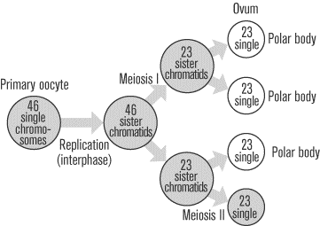
· Meiosis
o 2 Stages of Cell Division:
§ Meiosis I
· Homologous chromosomes come together and line up side by side (synopsis)
o Homologous chromosomes are pairs of chromosomes that look alike and carry genes for the same traits.
· This creates pairs of homologous chromosomes at the center of the spindle.
§ Meiosis II
· Centromeres divide and sister chromatids separate, becoming chromosomes that are distributed to daughter nuclei.
· The daughter cells mature into gametes that fuse during fertilization.
o Gametes: sex cells: sperm and eggs.
· Fertilization restores the diploid number of chromosomes in the zygote.
o Stages of meiosis
§ Meiosis I and Meiosis II, both have the same 4 stages of nuclear division as Mitosis.
o Prophase
o Metaphase
o Anaphase
o Telophase

o Significance of Meiosis
§ It is a part of gameogenesis, the production of the sperm and egg.
§ To keep the chromosome number constant from generation to generation.
§ The process results in genetic recombination, which ensures that offspring will be genetically different.
· Comparison of Meiosis and Mitosis
o Meiosis requires 2 nuclear divisions. Mitosis requires 1 nuclear division.
o Meiosis produces 4 daughter nuclei and following Cytokinesis there are 4 daughter cells. Mitosis results in 2 daughter cells.
o In meiosis 4 daughter cells are haploid and have half the chromosome number of the parent cell. In mitosis daughter cells have the same number of chromosomes as the parent cell (diploid).
o In meiosis daughter cells are not genetically identical to each other or to the parent cell. In mitosis the daughter cells are identical to each other and the parent cell.
o Meiosis occurs in the reproductive organs. Mitosis occurs in all tissue types during growth and repair.
· Spermatogenesis
o Is the production of sperm in males.
o All four daughter cells become sperm. Sperm has 23 chromosomes (the haploid number).

· Oogenesis
o Is the production of eggs in females.
· Chromosome Inheritance
o Normally an individual receives 22 pairs of autosomes and 2 sex chromosomes.
o Each pair of autosomes carries alleles for certain traits, the alleles can be different.
o Sometimes individuals are born with too many or too few autosomes or sex chromosomes.
§ This will usually happen during meiosis due to nondisjunction.
· Nondisjunction
o Occurs during meiosis I when both members of a homologous pair go into the same daughter cell.
o Or in meiosis II when a sister chromatin fails to separate and both daughter chromosomes go into the same gamete.
o It may occur during Oogenesis
o Normal development requires 2 kinds of each chromosome.
o Example of different chromosomal abnormalities
§ Down Syndrome
· Autosomal Trisomy
· Most common
· There will usually be 3 copies of chromosome 21.
· Karotyping is able to detect the defect.
· The genes that cause it are located on the bottom 3rd of chromosome 21.
§ Changes in sex chromosome #
· Results from inheriting too many or too few X or Y-chromosomes.
o Turner Syndrome (XO)
§ Has only 1 sex chromosome (X)
§ Persons do not go through puberty
§ Persons are of normal intelligence
o Klinefelter Syndrome (XXY)
§ Symptoms are very subtle
§ Speech and language delays
§ Will require assisted reproduction to father children
§ Receive testosterone supplementation at the start of puberty
o Poly X Female (XXX)
§ Has more than two X chromosomes and has extra barr bodies in the nucleus.
§ Tendency to be tall and thin
§ Delayed motor and language development.
o Jacobs Syndrome (XYY males)
§ Results from nondisjunction during spermatogenesis.
§ Taller than average
§ Persistent acne
§ Speech and reading problems.
· Changes in Chromosome structure
o Another type of chromosomes mutation when the chromosomes “break” and do not rejoin in the same pattern.
o Types include:
§ Deletion: the loss of a chromosomes piece.
§ Duplication: when a chromosomal segment is repeated in the same chromosome.
§ Inversion: when a piece of chromosome breaks loose and then rejoins in the reversed direction.
§ Translocation: the exchange of chromosome pieces between nonhomologous pairs.
o Human Syndromes
§ Deletion Syndromes
· Williams Syndromes
o Chromosome 7 loses a tiny end piece.
· Cru du chat (cats Cry)
o Chromosome 5 is missing an end piece.
· Translocation Syndromes
o The chromosome exchange breaks an allele into 2 piece; causing 1 of the translocated chromosomes to have only 1 copy of certain alleles and 3 copies of certain other alleles.
- DNA and RNA Structure and Function
- Structure of DNA
- Double helix, 2 strands that spiral.
- Each strand is a polynucleotide
- Polynucleotide: molecule of 3 subunits
- Phosphoric acid ( Phosphate)
- Pentose sugar (deoxyribose)
- Nitrogen containing base
- Phosphate- Sugar backbones are the support of the ladder.
- Paired Bases are the rungs held together by hydrogen bond.

- Replication of DNA
- The process of copying a DNA Helix.
- Before replication, the 2 strands that make up parental DNA are hydrogen bonded together.
- An enzyme “un zips” the double stranded DNA.
- New complimentary DNA nucleotides (always present in the nucleus) fit into place through complimentary base pairing.
- An enzyme seals any breaks in the phosphate-sugar backbone.
- The 2 double-helix molecules are identical to each other and to the original DNA molecule.

- A replication error that persists is a mutation
- Mutation: permanent change in the sequence of bases that may cause a change in the phenotype and introduce variability. The variability’s are what make you different.
- Structure and Function of RNA
- RNA is made of nucleotides containing the sugar ribose.
- Is single stranded
- In place of base thymine is uracil.
- Helper to DNA, allowing protein synthesis to occur according to the stored genetic information provided by DNA.
- Three types of RNA
- rRNA (ribosomal RNA)
- Joins with proteins made in the cytoplasm to form the subunits of ribosomes.
- mRNA (messenger RNA)
- Carries genetic information from DNA to ribosomes in the cytoplasm where protein synthesis occurs.
- tRNA (transfer RNA)
- Transfers amino acids to the ribosomes, where the amino acids are joined, forming a protein.
- Gene expression
- 2 Steps
- Transcription
- Translation
- Is possible only if the bases in DNA and mRNA code for amino acids, called genetic code.
- Genetic Code
- 4 bases stand for 1 amino acid
- Supplies 64 different triplets
- Each 3 letter unit of an mRNA molecule is a codon
- 61 triples correspond to a particular amino acid, remaining 3 are stop-codons which signal polypeptide termination.
- Transcription
- Occurs in the nucleus
- The DNA Helix opens to make a template.
- The mRNA transcript is made from the DNA template
- Translation
- Occurs outside the nucleus.
- The ribosomes do the translation.
- They glom onto mRNA and line up amino acids in a chain (polypeptide chain) according to mRNA sequence.
- Regulation of Gene Expression
- Cells differ by which genes are expressed.
- 4 primary levels of control
- Transcriptional Control
- In the nucleus
- The organization of chromatin and use of transcription factors that start transcription regulate gene transcription.
- Post Transcriptional Control
- In the nucleus
- The amount of the gene expressed is controlled by how fast mature mRNA leaves the nucleus.
- Translational Control
- In the cytoplasm
- Occurs after mRNA leaves nucleus and before there is a protein product.
- Post Translational Control
- In the cytoplasm
- Occurs after protein synthesis.
- Characteristics of Cancer Cells
· They lack differentiation
o Cells are non-specialized, they do not contribute to the functioning of body parts
o They look distinctly different.
· Abnormal Nuclei
o The nuclei are enlarged.
o It may have an abnormal number of chromosomes.
o The chromosomes are abnormal.
§ Portions may be deleted or duplicated
§ Gene amplification occurs (extra copies of specific genes)
o They do not go through aptopsis-programmed cell death.
· Unlimited Replicative Potential
o Cells are immortal and keep on dividing for an unlimited number of times (normal cells divide 60-70 times then die)
o The gene that codes for telomerase is turned on.
§ Telomerase: continually rebuilds the telomeres in cancer cells, remaining at a constant length (normal cells get shorter after each division and eventually die).
· Form Tumors
o Cells pile on top of one another and grow in multiple layers forming a tumor.
o Benign tumor- is encapsulated and will never invade adjacent tissues.
· No need for growth factors
o Cells divide when the stimulatory growth factors are not present.
o Cells do not respond to inhibitory growth factors.
· Cells gradually become abnormal
o Carcinogenesis- the development of Cancer
o 3 phases
§ Initiation: the cell undergoes mutation, causing it to divide repeatedly
§ Promotion: the tumor develops; tumor cells continue to divide, undergoing mutations.
§ Progression: One cell mutates to give it a selective advantage over other cells; the process is repeated giving the cells the ability to invade surrounding tissues.
· Undergo Angiogenesis and Metastasis
o Angiogenesis: formation of new blood vessels
§ Brings the tumor nutrients and oxygen to grow.
o Metastasis: is when cancer cells found in nearby lymphnodes, causes new tumors far from the 1st tumor.

· Cancer is a Genetic Disease
o Repeated cell cycles are caused by mutation of 2 types of genes.
§ Proto-oncogenes: code for proteins that promote the cell cycle and prevent apoptosis.
§ Tumor-suppressor genes- code for proteins that inhibit the cell cycle and promote apoptosis.
o Proto-Oncogenes -Oncogenes
§ Proto-oncogenes turn into oncogenes
§ Oncogenes are the result of mutation of proto-oncogenes.
§ Oncogenes are an over functioning proto-oncogene.
§ A gain of function mutation
o Tumor Suppressor Gene becomes inactive
§ Mutations cause the gene to no longer inhibit the cell cycle nor promote apoptosis.
§ Loss of function mutation
· Types of Cancer
o Oncology is the study of cancer.
o Tumors are classified according to place of origin.
§ Carcinomas- in the epithelial tissue
§ Sarcomas- in the muscle and connective tissue.
§ Leukemia’s- in the blood.
§ Lymphomas- in the lymphatic tissue.
· Causes of cancer
o Causes
§ Heredity
§ Environmental Carcinogens
· Radiation
o Uv light
o Radon gas
o X-rays
o Nuclear fuel
· Organic chemicals
o Tobacco smoke
o Pollutants
§ Asbestos, nickel, cadmium, uranium
· Viruses
o Hepatitis B and C
o Epstein-Barr Virus
o Human Papillomavirus
o Diagnosis of Cancer
§ Seven warning signs
· Change in bowel of bladder habits
· A sore that does not heal
· Unusual bleeding of discharge
· Thickening or lump in breast or other places
· Indigestion or difficulty swallowing
· Obvious change in wart or mole
· Nagging cough or hoarseness
§ Routine screening tests
· Self-examination followed by physician examination
· ABCD’s of melanoma
o A- Asymmetry, one half of mole does not look like the other half.
o B- Border, irregular scalloped or poorly circumscribed border.
o C- Color, varied from one area to another; shades of tan, brown, black, or sometimes white, red, or blue.
o D- Diameter, larger than 6mm.

· Tumor marker test
o Blood test for tumor antigens/ antibodies
· Genetic Tests
o Testing for genetic mutations in proto-oncogenes and tumor suppressor genes.
o Treatments of cancer
§ Surgery- for a specific location
§ Radiation Therapy- for a specific location
§ Chemotherapy- for the entire body
§ Bone Marrow Transplants
§ Genotypes and Phenotypes
o Genotype
§ Alleles: the alternative forms of a gene having the same position (Locus) on a pair of chromosomes and affecting the same trait.
§ Dominant allele: is assigned a upper case letter
§ Recessive Allele: assigned a lower case letter.
§ Occur in pairs, two alleles per trait
§ One of each pair of alleles is inherited from each parent.
§ Homozygous Dominant: 2 dominant alleles
§ Homozygous Recessive: 2 recessive alleles
§ Heterozygous: 1 dominant 1 recessive allele
o Phenotype
§ The physical appearance of the individual
§ Can be any characteristic of the individual.
§ One- and Two- Trait Inheritance
o One-Trait Crosses
§ The inheritance of only one set of alleles
o Two-Trait Crosses
§ The inheritance of two sets of alleles
o For both types of crosses it is necessary to determine the gametes of both parents.
§ Forming the Gametes
o Gametes are the same as alleles
o Gametes can carry one allele for each trait
o If an individual carries alleles “EE” all the gametes will carry an E
o If an individual carries an “Ee” half the gametes will carry an “E” and half will carry an “e”.
§ Autosomal Recessive Disorder
o Inheritance of 2 recessive alleles is required before the disorder will appear.
§ Autosomal Dominant Disorders
o Inheritance of only 1 dominant allele is necessary for the disorder to appear.
§ Beyond simple Inheritance Patterns
o In some patterns of inheritance the alleles are not just dominant or recessive.
o Polygenic inheritance
§ Polygenic traits are controlled by more than one set of alleles. The dominant alleles have an additive effect on the phenotype.
o Incomplete Dominance and Codominance
§ Incomplete Dominance: the heterozygote is intermediate between the 2 homozygotes
§ Codominance: both dominant alleles are expressed equally.
§ Multiple Allele Inheritance
o Every individual has 2 out of 3 possible alleles
Thursday, February 14, 2008
Microscope Lab

This is a image of cheek cells under 40X magnification.
The Microscope
History of the Microscope
The first microscope made was by Hans and Zacharias Janssen around 1595in Middleburg, Holland. The compound microscope that they made was a tube with lenses at both ends. The magnification range of this scope ranged from 3X to 9X.
Robert Hooke made and improvement to the compound microscope around 1600. He discovered the features of plant tissue under the microscope and called his findings plant cells.
Anton van Leeuwenhoek built the best simple microscopes in his time 1632-1723. He was the fist person to describe bacteria, and helped to prove the theory of blood circulation.
Types of Microscopes
There are four types of microscopes: The Compound Microscope, Dissection Microscope, Scanning Electron Microscope, and the Transmission electron Microscope. The compound microscope is light illuminated. The image seen is two-dimensional. It is the most commonly used out of the different microscopes. It has a high magnification and a low resolution. The dissection microscope is light illuminated. The image seen is 3-dimensional. It has a low magnification; you are not able to see individual cells. The scanning electron microscope uses electron illumination. The image seen is 3-dimensional. It has high magnification and resolution. The transmission electron microscope is electron illuminated. The image seen is 2-dimensional. It has high magnification and resolution.
How to Use a Microscope
1. Turn on the light
2. Adjust the rheostat dial to 10; the dial moves left and right.
3. Adjust the iris diaphragm to change the amount of light coming through the aperture, the iris lever moves left to right.
4. Put your slide on the stage using the stage clips.
5. Make sure the 4X lens is in place; the dial moves left and right.
6. Move the stage left and right and up and down until the specimen is centered over the aperture.
7. Use the course focus dial to move the stage as high as it will go, turn the dial up.
8. After you have done these steps you now need to look through the oculars.
9. You need to adjust the oculars until the 2 circles of light are one circle.
10. You will then need to adjust the coarse focus dial down until you see the specimen (only use the coarse dial when on low powered scope)
11. After you have adjusted the coarse dial and see the specimen, adjust the fine focus knob to sharpen the image.
12. Re-center the specimen if needed.
13. Adjust the iris diaphragm to adjust the amount of light.
14. Once the specimen is in focus you can then move to the next higher-powered lens.
15. You will then repeat steps 11-13 to get the specimen in focus.
16. Repeat steps 11-15 for all remaining higher-powered lenses.


Subscribe to:
Posts (Atom)
