Regarding My Work
1. What were the three aspects of the assignments I've submitted that I am most proud of?
Three aspects of the assignments i'm most proud of are how they are organized, how the pictures came out for the lab project, and how complete the compendium review is.
2. What two aspects of my submitted assignments do I believe could have used some improvement?
Two aspects that could have used some improvement are the ethical issues essay, and I missed some parts that needed to be included in the unit lab.
3. What do I believe my overall grade should be for this unit?
I dont know hopefully a B
4. How could I perform better in the next unit?
Make sure I have a better understanding of the unit.
Regarding The Unit
At what moment during this unit did you feel most engaged with the course?
During the lab project
At what moment unit did you feel most distanced from the course?
Doing the compendium review.
What action that anyone (teacher or student) took during this unit that find most affirming and helpful?
None that I can think of.
What action that anyone (teacher or student) took during this unit did you find most puzzling or confusing?
What about this unit surprised you the most? (This could be something about your own reactions to the course, something that someone did, or anything else that occurs to you.)
How involved the process is to contract a muscle.
Monday, April 14, 2008
Lab Project Unit Three
This is a model of how the muscles and bones work together to create movement. You will first see how the muscle and bone are attached and when the muscle is contracted. The remaining models show what happens to the muscle fibers when a muscle contracts.
Limb Parts and the Model Representations
Bones: Card Board
Muscle: Toilet Paper
Actin Filaments: Cheerios
Myosin: Masking Tape
Cross-bridge: Orange Dots on Myosin
Z-Line: Black Line
Myosin Binding Site: Black Dots on Cheerios
Myosin Head: Card Board Strips
Tropomyosin: Black String
Troponin: Corn Kernels
Calcium: Marshmallow


The models shows how the actin filaments look when they are relaxed and contracted. You also see how calcium causes the actin filaments and the myosin to bind, which causes the actin filaments to contract or shorten. This reaction is what causes a muscle to shorten and contract, creating the ability to move our bones.
Unit 3 Topic 2 Online Lab
How Do Your Muscles Work?
1. Effect of Temperature on Muscle Action
Temperature Number of Fists
Normal 34
Freezing 20
2.Effect of Fatigue on Muscle Action
Trial # of Squeezes in 20 Seconds
1 41
2 40
3 36
4 34
5 34
6 33
7 33
8 31
9 30
10 30
ANALYSIS OF DATA:
1.What are the three changes you observed in a muscle while it is working
(contracted)?
The muscle gets hard, shorter, and has a bigger circumference.
2.What effect did the cold temperature have on the action of your hand muscles?
Explain.
It made muscle contraction slower.

Attempts Number
4. What effect did fatigue have on the action of your hand muscles? Explain.
As my muscles got fatigued it was harder to move them, and they moved slower, the more tired they got.
As the energy supply is depleted in the cells your muscles become fatigued and quits responding even when you are trying to make them work.
Ethical Issue: Exercise…is the concept working?
How often do you exercise? What do you consider exercise? Exercise is any activity that makes your body move. The different types of exercise are put into 3 categories: Aerobic, Strength, and flexibility. Our environment has evolved so that exercising is not part of our daily routine. There are several factors that are believed to be the cause of this.
The way that our communities are built is one reason why we do not exercise enough. With the convenience of drive thru windows and fast food there is no need to get out of the car and produce and kind of physical activity. These conveniences allow us to be as inactive as possible. We have the ability to have what we need delivered to our homes allowing us to be even less active.
Older communities were built with no sidewalks or bike paths. Making exercise dangerous because of passing vehicles. There were not many parks built so it was inconvenient to go to the park and exercise.
Home entertainment that is advertised heavily is another reason why we are so inactive. DVD’s, video games, even television does nothing to promote being active. People would rather sit and watch a movie than go hiking. Kids are learning to be inactive at an early age, which will make obesity rates go up. If they get into the habit of not exercising when they are young it will be hard for them to change the habit when they are older.
The convenience of drive thru windows, not enough safe places to exercise and home entertainment are a big cause of why we are no longer active. We want convenience and exercise has become an inconvenience.
People can exercise in many ways. Being active by walking to the mailbox instead of driving or going dancing once a week is considered exercise. I think that if we do not educate our children and control the amount of video games and television that they watch the obesity epidemic will get worse. We need to continue to build more communities with more parks, bike paths and sidewalks. We need to show our children the many different ways of exercising that can be incorporated into our everyday routine, like taking the dog for a walk or playing a game of basket ball with their dad.
Saturday, April 12, 2008


QUESTIONS ABOUT LEECH NEUROPHYSIOLOGY LAB:
1. What is the electrode measuring?
The electrode is measuring the potential difference across the neurons membrane
2. Why use leeches in neurophysiology experiments?
A leech has a simple nervous system, which makes it easier to understand and the nervous system also obey’s many of the same rules as our complex one’s.
3. What is the difference between a sensory and a motor neuron?
A sensory neuron takes nerve impulses from a sensory receptor to the CNS, a motor neuron takes nerve impulses away from the CNS to an effector.
4. Do you think a leech experiences pain? What is pain?
Yes, because it has a nervous system.
5. What were the two most interesting things about doing this lab?
Learning about the leech, and seeing how it was dissected.
6. Anything you found confusing or didn't like about the lab?
no
Friday, April 11, 2008
• Topic One chapters 13 and 14
• Chapter 13
• Overview of the nervous system

o There are 2 main parts to the nervous system
Central Nervous System (CNS)
• Brain
• Spinal Cord
Peripheral Nervous System
• Nerves outside the CNS
o There are 3 specific functions
Receiving sensory input
CNS performs integration
CNS generates motor output
o Nervous Tissue
Contains 2 types of cells

• Chapter 13
• Overview of the nervous system
o There are 2 main parts to the nervous system
Central Nervous System (CNS)
• Brain
• Spinal Cord
Peripheral Nervous System
• Nerves outside the CNS
o There are 3 specific functions
Receiving sensory input
CNS performs integration
CNS generates motor output
o Nervous Tissue
Contains 2 types of cells
• Compendium Review Unit #3
• Neurons
o Cells that transmit nerve impulses between parts of the nervous system
• Neuroglia
o Support and nourish neurons
o Neuron Structure
3 types of neurons
• Sensory neurons
o Takes nerve impulses (messages) from a sensory receptor to the CNS
Sensory receptor is a special structure that detects changes in the environment.
• Interneuron
o Is completely within the CNS, they receive input from sensory neurons and from other interneurons in the CNS.
o After receiving the information they sum it all up before translating it to motor neurons
• Motor Neuron
o Takes nerve impulses away from the CNS to an effector (muscle fiber or gland)
Effectors carry out our responses to environmental changes, external and internal.
o Myelin Sheath

Is the protective cover over the axons.
Is formed by neuroglia cells called Schwann Cells
• Schwann Cells contain myelin in their plasma membrane
• Develops when these cells wrap themselves around an axon many times.
• Each neuroglia cell only covers a part of the axon, the gaps where there is no cover is called nodes of Ranvier.
Long Axons have a myelin sheath, but short axons do not.
Myelin gives nerve fibers their white glistening appearance and is also an insulator.
The myelin sheath is important to nerve regeneration within the PNS.
• If an axon is severed, the myelin sheath remains and serves as a passageway for new fiber growth.
o The Nerve Impulse
Nerve impulses convey information within the nervous system.
• Nerve impulse is studied by using excised axons and a voltmeter to measure voltage.
o The voltmeter allows measurement of the potential difference between 2 sides of the axonal membrane
Resting potential
• Is when the axon is not conducting an impulse.
• The inside of the neuron is more negative than the outside
• Resting potential correlates to a difference in ion distribution on either side of the axonal membrane
o More Na+(sodium ions) outside the axon
o More K+ (potassium ions) inside the axon
o Sodium-Potassium Pump created the unequal distribution of these ions.
Actively transports Na+ out of the axon
Actively transports K+ into the axon
The membrane is permeable to K+ but not to Na+, causing there to always be more positive ions outside the membrane than inside.
Action Potential
• Is a rapid change in polarity across an axonal membrane as the nerve impulse occurs.
• If a stimulus causes the axonal membrane to depolarize to a certain level, called threshold, an action potential occurs in an all-or-none manner.
• Action potential requires 2 types of gated channel proteins in the membrane
o Sodium Gates Open
When action potential occurs, gates of sodium channels open first
Na+ flows into the axon.
This causes a depolarization because the charge inside the axon changes from negative to positive.
o Potassium Gates Open
Second, the gates of the potassium channels open
K+ flows to outside the axon.
This causes a repolarization because the inside of the axon resumes a negative charge as K+ exits he axon.
Visualizing an Action Potential
• After an action potential has passed by, the sodium-potassium pump restores the resting potential by moving the potassium back to the inside and sodium back to the outside.
o Propagation of an Action Potential
Action potentials are self-propagating; each action potential generates another along the length of an axon.
When an axon does not have a myelin sheath, the action potential at one locale stimulates an adjacent part of the axon’s membrane to produce an action potential.
In axons that have a myelin sheath, an action potential at one ranvier node causes an action potential at the next node.
o The Synapse
Every axon branches into many fine endings, each is tipped by a small swelling called an axon terminal.
Each axon terminal lies very close to either the dendrite or the cell body of another neuron
This region of close proximity is called a synapse
• At a synapse, a small gap called the synaptic cleft separates the sending neuron from the receiving neuron.
Transmission across a synapse is carried out by molecules called neurotransmitters.
These events occur:
• Nerve impulses traveling along an axon reach an axon terminal.
• Calcium ions enter the terminal, and they stimulate synaptic vesicles to merge with the sending membrane
• Neurotransmitter molecules are released into the synaptic cleft, and they diffuse across the cleft to the receiving membrane, where they bind with specific receptor proteins.
Neurotransmitter Molecules
• More than 100 substances are known or suspected to be neurotransmitters, some common ones are:
o Acetylcholine and norepinephrine
Active in both the CNS and PNS
Norepinephrine excites smooth muscle in the CNS
Acetylcholine in the PNS; excites skeletal muscle and inhibits cardiac muscle.
o GABA (gamma aminobutyric acid)
Is an abundant inhibitory neurotransmitter in the CNS
o Serotonin
Involved in thermoregulation, sleeping, emotions, and perception
• The Central Nervous System
o The spinal cord and the brain make up the CNS
o Sensory information is received and motor control is initiated.
o The Spinal Cord

Extends from the base of the brain through a large opening in the skull called the foramen magnum and into the vertebral canal formed by openings in the vertebrae.
Structure of the spinal cord
• Spinal nerves project from the cord between the vertebrae that make up the vertebral column.
• Central canal contains cerebrospinal fluid, along with the meninges that protect the spinal cord.
• The gray matter is centrally located and shaped like the letter H.
o This is where portions of the sensory neurons and motor neurons are found, interneurons that communicate with these 2 types of neurons are also found there.
• Dorsal root contains sensory fibers entering the gray matter
• Ventral roots contains motor fibers exiting the gray matter
• The dorsal and ventral roots join before the spinal nerve leaves the vertebral canal as a mixed nerve
• White matter occurs in areas around the gray matter
o It contains ascending tracts taking information to the brain and descending tracts taking info from the brain.
Functions of the Spinal Cord
• Provides a means of communication between the brain and the peripheral nerves that leave the cord.
• Sensory receptors generate nerve impulses that pass through sensory fibers to the spinal cord and up ascending tracts to the brain.
• Gate Control Theory of Pain:
o The tracts in the spinal cord have ”gates,” and these “gates” control the flow of pain messages from the peripheral nerves to the brain.
o Depending on how the gates process a pain signal, the pain message can be allowed to pass directly to the brain or it can be prevented from reaching the brain.
o When the brain initiates voluntarily control over our limbs, motor impulses originating in the brain pass down descending tracts to the spinal cord and out to our muscles by way of motor fibers
o If the spinal cord is severed, we suffer a loss of sensation and a loss of voluntary control.
If the cut occurs in the thoracic region, the lower body and legs are paralyzed, known as paraplegia.
If the injury is in the neck region, all four limbs are usually affected, known as quadriplegia.
o Reflex Actions
A stimulus causes sensory receptors to generate nerve impulses that travel in sensory axons to the spinal cord
Interneurons integrate the incoming data and relay signals to motor neurons
A response to the stimulus occurs when motor axons cause skeletal muscles to contract.
Each interneuron in the spinal cord has synapses with many other neurons, they send signals to several other interneurons and motor neurons.
o The Brain
The Cerebrum
• Also referred to as the Telencephalon
• Is the larges part of the brain.
• The last center to receive sensory input and carry out integration before commanding voluntary motor responses
• It communicates with and coordinates the activities of the other parts of the brain.
• Cerebral Hemispheres
o 2 halves
Left Cerebral Hemisphere
Right Cerebral Hemisphere
o A deep groove called the longitudinal fissure divides the left and right cerebral hemispheres.
o Shallow grooves called sulci divide each hemisphere into lobes
Frontal Lobe
• Is the most ventral of the lobes
• Located directly behind the forehead
Parietal lobe
• Dorsal to the frontal lobe
Occipital Lobe
• Dorsal to the parietal lobe
• Located at the real of the head
Temporal lobe
• Inferior to the front and parietal lobes
• At the temple and the ear
• The Cerebral Cortex
o Is a thin but highly convoluted outer layer of gray matter that covers the cerebral hemispheres.
o Is the region of the brain that accounts for:
Sensation
Voluntary movement
All the thought processes we associate with consciousness
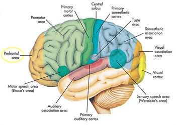
• Primary Motor and Sensory Areas of the Cortex
o Primary Motor Area
Located in the frontal lobe just before the central sulcus.
Voluntary commands to skeletal muscles begin in the primary motor area, each part of the body is controlled by a certain section
o Primary Somatosensory Area
Is located dorsal to the central sulcus in the parietal lobe
Sensory information from the skin and skeletal muscles arrives here, where each part of the body is sequentially represented
• Primary taste area
o Accounts for taste sensations
• Primary visual area
o In the occipital lobe receives information from our eyes
• Primary auditory area
o In the temporal lobe receives information from our ears
• Primary olfactory area
o Is for smell, located in the temporal lobe
o Association Areas
Located in places where integration occurs
Ventral to the primary motor area is a pre-motor area
• The pre-motor area organizes motor functions for skilled motor activities, such as walking and talking at the same time
• The primary motor area then sends signals to the cerebellum, which integrates them.
o Processing Centers
Processing centers of the cortex receive information from the other association areas and perform higher-level analytical functions
The prefrontal area, an association area in the frontal lobe, receives information from other association areas and uses this information to reason and plan our actions
o Central White Matter
Most of the remaining cerebrum is composed of white matter
The Diencephalon
• Is a region that encircles the third ventricle
• Hypothalamus and the thalamus are located here
o Hypothalamus forms the floor of the third ventricle
Integrating center that regulates hunger, sleep, thirst, body temperature, and water balance,
Controls the pituitary gland, is the link between the nervous and endocrine system
o Thalamus consists of 2 masses of gray matter located in the sides and roof of the third ventricle.
Is on the receiving end for all sensory input except smell
Visual, auditory, and somatosensory information arrives at the thalamus via the cranial nerves and tracts from the spinal cord.
The thalamus integrates this information and sends it on to the appropriate portions of the cerebrum.
The Cerebellum
• Lies under the occipital lobe of the cerebrum and is separated from the brain stem by the fourth ventricle
• Made up of 2 portions that are joined by a narrow median portion
• Receives sensory input from the eyes, ears, joints, and muscles about the present position of body parts, and receives motor output from the cerebral cortex about where these parts should be located.
• After integrating this information, the cerebellum sends motor impulses by way of the brain stem to the skeletal muscles.
The Brain Stem
• Contains the midbrain, the pons, and the medulla oblongata
o Midbrain
Acts as a relay station for tracts passing between the cerebrum and the spinal cord or cerebellum.
Has reflex centers for visual, auditory, and tactile responses
o Pons
Contains bundles of axons traveling between the cerebellum and the rest of the CNS.
Functions with the medulla oblongata to regulate breathing rate and has reflex centers concerned with head movements in response to visual and auditory stimuli
o Medulla Oblongata
Contains a number of reflex centers for regulating heartbeat, breathing, and blood pressure.
Contains reflex centers for vomiting, coughing, sneezing, hiccupping, and swallowing.
• The Limbic System and Higher Mental Functions
o Limbic System
An evolutionary ancient group of linked structures deep within the cerebrum that is a functional grouping rather than an anatomical one
It blends primitive emotions and higher mental functions into a united whole.
There are two structures within the limbic system
• The amygdala
o Can cause experiences to have emotional overtones
o Creates the sensation of fear.
• The Hippocampus
o Believed to play a crucial role in learning and memory
Acts as an information gateway during the learning process, determining what information about the world is to be sent to memory, and how the information is to be encoded and stored by other regions in the brain
o Higher Mental Functions
Types of memory
• Short-term memory
• Long-term memory
• Semantic memory: number, words
• Episodic memory: persons, events
• Skill Memory: involved in performing motor activities
Long-Term Memory Storage and Retrieval
• Long-term memories are apparently stored in bits and pieces throughout the sensory association areas of the cerebral cortex.
• The Peripheral Nervous System
o Nerves that lie outside the CNS
o 2 types
Cranial Nerves
• Attached to the brain
• 12 pairs
Spinal Nerves
• Emerge in 31 pairs from either side of the spinal cord.
o Somatic System
Nerves in the somatic system serve the skin, skeletal muscles, and tendons.
Includes nerves that take sensory information from external sensory receptors to the CNS and motor commands away from the CNS to the skeletal muscles.
Automatic responses to a stimulus in the somatic system are called reflexes
• The Reflex Arc
o The path of a reflex that involves only the spinal cord.
o Sensory receptors in the skin generate nerve impulses that move along sensory fibers through the dorsal-root ganglia toward the spinal cord.
o Sensory neurons that enter the cord dorsally pass signals on to many interneurons.
o Nerves impulses travel along motor fibers to an effector, which brings about a response to the stimulus.
o The whole series of responses occurs because some of the interneurons involved carry nerve impulses to the brain.
o Autonomic System
Located in the PNS
Regulates the activity of cardiac and smooth muscles and glands
Divided into the sympathetic and parasympathetic divisions.
• Shared features are:
o They function automatically and in an involuntary manner
o They innervate all internal organs
o They utilize 2 neurons and one ganglion for each impulse
• Sympathetic division
o Important to fight or flight situations
o Accelerates the heart beat and dilates the bronchi
• Parasympathetic Division
o Promotes all the internal responses we associate with a relaxed state
• Chapter 14
• Sensory Receptors and Sensations

o Sensory Receptors
Are dendrites that are specialized to detect certain types of stimuli
Exteroceptors
• Sensory receptors that detect stimuli from outside the body
• Ex: those that result in taste, smell, vision, hearing, and equilibrium
• Are not directly involved in homeostasis and continually send messages to the central nervous system regarding environmental conditions
Interoceptors
• Receive stimuli from inside the body
• Ex: pressoreceptors respond to changes in blood pressure, osmoreceptorsdetect changes in water-balance
• Are directly involved in homeostasis and are regulated by a negative feedback mechanism
o Types of Sensory Receptors
Chemoreceptors
• Respond to chemical substances in the immediate vicinity
• Ex: taste, smell,
• Pain Receptors
o Are a type of chemoreceptor, they are naked dendrites that respond to chemicals released by damaged tissues
Photoreceptors
• Respond to light energy
• Our eyes contain photoreceptors that are sensitive to light rays and provide us with a sense of vision.
Mechanoreceptors
• Are stimulated by mechanical forces, which most often result in pressure of some sort.
Thermoreceptors
• Are located in the hypothalamus and skin and are stimulated by changes in temperature.
o Warmth receptors respond when temperatures rise
o Cold receptors respond when temperatures lower
o How Sensation Occurs
Sensory receptors respond to environmental stimuli by generating nerve impulses.
When nerve impulses arrive at the cerebral cortex of the brain, sensation, which is the conscious perception of stimuli, occurs.
All sensory receptors initiate nerve impulses; the sensation that results depends on the part of the brain receiving the nerve impulses
• Proprioceptors and Cutaneous Receptors
o Proprioceptors
Are mechanoreceptors involved in reflex actions that maintain muscle tone, and thereby the body’s equilibrium and posture.
They help us know the position of our limbs in space by detecting the degree of muscle relaxation, the stretch of tendons, and the movement of ligaments.
Muscle spindles increase the degree of muscle contraction
Golgi tendon organs decrease the degree of muscle contraction
o Cutaneous Receptors
Are located in the dermis, making the skin sensitive to touch, pressure, pain, and temperature.
There are 3 types of receptors that are sensitive to fine touch
• Meissner corpuscles
o Along with Krause end bulbs are concentrated in the fingertips, the palms, the lips, the tongue, the nipples, the penis, and the clitoris.
• Merkel disks
o Located where the epidermis meets the dermis
• Root Hair Plexus
o Is a free nerve ending that winds around the base of a hair follicle and fires if the hair is touched.
2 types of receptors that are sensitive to pressure
• Pacinian corpuscles
o Are onion shaped receptors that lie deep inside the dermis.
• Ruffini Endings
o Are encapsulated by sheaths of connective tissue and contain lacy networks of nerve fibers.
o Pain Receptors
Nociceptors
• Pain receptors for internal organs that are sensitive to chemicals released by damaged tissues.
• When inflammation occurs, because of mechanical, thermal, or electrical stimuli or toxic substances, cells release chemicals that stimulate pain receptors.
• Senses of Taste and Smell
o Taste and smell are chemical senses because their receptors are sensitive to molecules in the food we eat and the air we breathe.
o Chemoreceptors are plasma membrane receptors that bind to particular molecules. They are divided into 2 types:
Those that respond to distant stimuli
• Olfactory cells act from a distance
Those that respond to direct stimuli
• Taste cells act directly
o Sense of Taste
Taste buds are primarily located on the tongue
Four Primary types of taste
• Sweet
• Sour
• Salty
• Bitter
• A fifth taste called umami may exist for certain flavors of:
o Cheese
o Beef broth
o Some sea food
Certain regions of the tongue are more sensitive to different types of taste.
How the Brain Receives Taste Information
• Taste buds open at a taste pore.
• They have supporting cells and a number of elongated taste cells that end in microvilli.
• When molecules bind to receptor proteins of microvilli nerve impulses are generated in sensory nerve fibers that go to the brain.
• When they reach the gustatory (taste) cortex, they are interpreted as particular tastes.
o Sense of Smell
80-90% of what we perceive as taste is actually due to the sense of smell.
Our sense of smell depends on 10-20 million olfactory cells located within olfactory epithelium high in the roof of the nasal cavity.
How the Brain receives Odor Information
• Each olfactory cell has one out of several hundred different types of receptor proteins.
• Nerve fibers from like olfactory cells lead to the same neuron in the olfactory bulb, an extension of the brain.
• An odor contains many odor molecules, which activate a characteristic combination of receptor proteins.
• An odor’s signature in the olfactory bulb is determined by which neurons are stimulated.
• When neurons communicate this information via the olfactory tract to the olfactory areas of the cerebral cortex.
• Sense of Vision
o Anatomy and Physiology of the Eye

Is an elongated sphere about 2.5 cm in diameter
Has 3 Layers
• The Sclera
o The outer layer
o Is white and fibrous
o Except for the cornea, which is made of transparent collagen fibers
• The Choroid
o The middle, thin darkly pigmented layer
o Is vascular and absorbs stray light rays that photoreceptors have not absorbed.
o Toward the front it becomes the iris.
Regulates the size of the pupil
Pupil is a hole in the center of the iris that lets light into the eyeball.
o Behind the iris the choroids thickens and becomes ciliary body
Controls the shape of the lens for near and far vision
o The lens, divides the eye into 2 compartments
The anterior compartment
• Is filled with a clear watery fluid called aqueous humor
• Located in front of the lens
Posterior Compartment
• Located behind the lens
• Is filled with vitreous humor, a clear gelatinous material
• The Retina
o Located in the posterior compartment
o Contains photoreceptors called
Rod Cell
• Very sensitive to light
• Do not see color
Cone Cells
• Require bright light
• Sensitive to different wavelengths of light, giving us the ability to distinguish colors.
o Fovea centralis
Region where cone cells are packed densely
Light is normally focused here when we directly look at an object
o Optic Nerve
Formed from sensory fibers in the retina
Takes nerve impulses to the visual cortex
Function of the Lens
• The cornea, assisted by the lens and humors, focuses images on the retina.
• Visual Accommodation
o Occurs for close vision
o The lens rounds up, in order to bring the image to focus on the retina.
o The ciliary muscle contracts, releasing tension on the suspensory ligaments, the lens rounds up due to the natural elasticity.
• Sense of Hearing
o The ear has 2 sensory functions:
Hearing
Balance
The sensory receptors for both are located in the inner ear
Each consists of hair cells with stereocilia that are sensitive to mechanical stimulation. They are mechanoreceptors
o Anatomy and Physiology of the Ear

The ear has 3 divisions
• Outer
o Outer ear consists of the pinna and the auditory canal
• Middle
o Begins at the tympanic membrane and ends at a bony wall containing two small openings covered by membranes
These openings are called the:
• Oval window
• Round window
o Three small bones are found between the tympanic membrane and the oval window. They are called the ossicles
Malleus (hammer)
Incus (anvil)
Stapes (stirrup)
o The Eustachian Tube is an auditory tube that extends from the middle ear to the nasopharynx, it permits equalization of air pressure
• Inner
o Is filled with fluid
o Has three areas:
Semicircular canals
• Concerned with equilibrium
Vestibule
• Concerned with equilibrium
Cochlea
• Concerned with hearing
o Auditory Pathway to the Brain
Through the Auditory Canal and Middle Ear
• The process begins when sound waves enter the auditory canal.
• When a large number of waves strike the tympanic membrane, it moves back and forth ever so slightly.
• The malleus then takes the pressure from the inner surface of the tympanic membrane and passes it to the stapes.
• The stapes strikes the membrane of the oval window, causing it to vibrate; the pressure is passed to the fluid within the cochlea.
From the Cochlea to the Auditory Cortex.
• Cochlea has 3 canals
o The cochlear canal
o The vestibular canal
o The tympanic canal
• When the stapes strikes the membrane of the oval window, pressure waves move from the vestibular canal to the tympanic canal across the basilar membrane.
• The basilar membrane moves up and down, and the stereocilia of the hair cells embedded in the tectorial membrane bend.
• Nerve impulses begin in the cochlear nerve and travel to the brain.
• When they reach the auditory cortex in the temporal lobe, they are interpreted as a sound.
• Sense of Equilibrium
o Rotational Equilibrium Pathway
Mechanoreceptors in the semicircular canals detect rotational and angular movement of the head.
The three semicircular canals are arranged so that there is one in each dimension of space
The base of each of the three canals, called the ampulla, is slightly enlarged.
Little hair cells, whose stereocilia are embedded within a gelatinous material cupula, are found within the ampullae.
Because of the way the semicircular canals are arranged, each ampulla responds to head rotation in a different plane of space.
As fluid within a semicircular canal flows over and displaces a cupula, the stereocilia of the hair cells bend, and the pattern of impulses carried by the vestibular nerve to the brain changes.
The brain uses information from the hair cells within ampulla of the semicircular canals to maintain equilibrium through appropriate motor output to various skeletal muscles that can right our present position in space as need be.
o Gravitational Equilibrium Pathway
The mechanoreceptors in the utricle and saccules detect movement of the head in the vertical or horizontal planes.
The utricle and saccules are two membraneous sacs located in the inner ear near the semicircular canals.
Both of these sacs contain little hair cells, whose stereocilia are embedded within a gelatinous material called an otolithic membrane. Calcium carbonate granules, otoliths, rest on this membrane.
When the body is still, the otoliths in the utricle and the saccules rest on the otolithic membrane above the hair cells
When the head bends or the body moves in the horizontal and vertical planes, the otoliths are displaced and the otolithic membrane sags, bending the stereocilia of the hair cells beneath.
If the stereocilia move toward largest stereocilium, called the kinocilium, nerve impulses increase in the vestibular nerve.
If the stereocilia move away from the kinocilium, nerve impulses decrease in the vestibular nerve.
The frequency of nerve impulses in the vestibular nerve indicates whether you are moving up or down.
These data reach the brain, which uses them to determine the direction of the movement of the head at the moment.
The brain uses this information to maintain gravitational equilibrium through appropriate motor output to various skeletal muscles that can right our present position in space as need be.
• Chapter 11
• Overview of Skeletal System

o Functions of the Skeleton
The skeleton supports the body.
• Bones of the legs support the entire body when we are standing.
• The bones of the pelvic girdle support the abdominal cavity.
The skeleton protects soft body parts.
• Bones of the skull protect the brain
• The rib cage protects the heart and lungs.
• The vertebrae protect the spinal cord.
The skeleton produces blood cells
• All bones in the fetus have red bone marrow that produces blood cells
• Only certain bones in adults produce blood cells.
The skeleton stores mineral and fat
The skeleton, along with the muscles, permits flexible body movement.
o Anatomy of a Long Bone
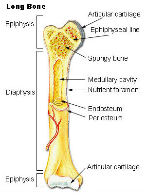
The diaphysis
• The shaft, or main portion of the bone
• Has a large medullary cavity
o Walls are composed of compact bone
o Lined with a thin, vascular membrane, and is filled with yellow bone marrow that stores fat.
The epiphyses
• Expanded region at the end of a long bone
• Composed largely of spongy bone that contains red bone marrow
• Coated with a thin layer of hyaline cartilage, called articular cartilage
Periosteum
• A layer of fibrous connective tissue covering the long bone
• Contains blood vessels, lymphatic vessels, and nerves
• Is continuous with ligaments and tendons that are connected to a bone.
Bone
• Compact Bone
o Highly organized and composed of tubular units called osteons.
• Spongy Bone
o Unorganized appearance
o It contains numerous thin plates called trabeculae, separated by unequal spaces
Cartilage
• Not as strong as bone
• More flexible than bones
• Has no nerves
• Made to pad joints where the stresses of movement are intense.
• Three types of cartilage
o Hyaline Cartilage
Firm and somewhat flexible
The matrix is uniform and glassy
Found at the ends of long bones, in the nose, at the end of the ribs, and in the larynx and trachea.
o Fibrocartilage
Stronger than hyaline
Able to withstand tension and pressure
Found in the disks located between the vertebrae and in the cartilage of the knee.
o Elastic cartilage
More flexible than hyaline cartilage
Found in the ear flaps and the epiglottis
• Fibrous connective tissue
o Contains rows of cells called fibroblasts separated by bundles of collagenous fibers
o Makes up the ligaments that connect bone to bone.
o Makes up the tendons that connect muscles to a bone at joints (articulations).
• Bone Growth, Remodeling, and Repair
o Several different types of bone cells are involved in bone growth, remodeling, and repair.
Osteoblasts
• Bone-forming cells
• They secrete the organic matrix of bone and promote the deposition of calcium salts into the matrix.
Osteocytes
• Mature bone cells
• Derived from osteoblasts
• Maintain the structure of bone
Osteoclasts
• Bone-absorbing cells
• They break down bone and assist in depositing calcium and phosphate in the blood.
o Bone Growth and Development
Ossification
• The formation of bone.
The bones of the skeleton form during embryonic development in 2 ways:
• Intramembranous Ossification
o Flat bones are examples of intramembranous Ossification.
o Bones develop between sheets of fibrous connective tissue.
o Cells derived from connective tissue cells become osteoblasts located in the ossification centers.
o Osteoblasts secrete the organic matrix of bone consisting of mucopolysaccharides and collagen fibrils.
o Calcification occurs when calcium salts are added to the organic matrix
o Osteoblasts promote calcification
o Ossification results in the trabeculae of spongy bone, spongy bone remains on the inside.
o Periosteum forms outside the spongy bone and osteoblasts, derived from the periosteum, carry out further ossification.
o Trabeculae form and fuse to become compact bone, which surrounds the spongy bone.
• Endochondral Ossification
o How most of the bones of the human skeleton are formed.
o Bone replaces cartilaginous models of the bones.
o Gradually, the cartilage is replaced by the calcified bone matrix that makes these bones capable of bearing weight.
• Epiphyseal Plates
o Have four layers
Resting Zone
• Layer nearest the epiphysis
• Where cartilage remains
Proliferating Zone
• Next to the resting zone
• Chondrocytes are producing new cartilage cells.
Degenerating Zone
• The third layer
• The cartilage cells are dying off
Ossification Zone
• Fourth layer
• Bone is forming
• Final Size of the Bones
o When epiphyseal plates close, bone length can no longer occur.
o The arms and legs of women close at about age 18.
o The arms and legs of men close at about age 20.
o Portions of other types of bones may continue until age 25.
o Hormones control the activity of the epiphyseal plate.
• Hormones Affect Bone Growth
o Several different hormones are involved in bone growth.
Growth Hormone (GH)
• Directly stimulates growth of the epiphyseal plate, and bone growth in general.
Thyroid Hormone
• GH would be ineffective if the metabolic activity of cells is not promoted.
• Thyroid Hormone promotes the metabolic activity of cells.
o Bone Remodeling and Its Role in Homeostasis
Bone Remodeling
• The process of bone renewal to keep bones strong.
• Allows the body to regulate the amount of calcium in the blood.
• The blood calcium level is critical because:
o If calcium concentration is too high, neurons and muscle cells no longer function
o If calcium concentrations fall too low, convulsions occur.
• Bones absorb excess calcium if level is too high.
• Bones release calcium if level is too low.
o Bone Repair
Is required after bone breaks or is fractured
Fracture repair takes several months in a series of 4 steps:
• Hematoma
o After a fracture blood escapes from ruptured blood vessels and forms a hematoma in the space between the ends of the broken bone
o Happens within 6-8 hours.
• Fibrocartilaginous Callus
o Tissue repair begins
o Fibrocartilaginous callus fills the space between the ends of the broken bone for about 3 weeks.
• Bony Callus
o Osteoblasts produce trabeculae of spongy bone and convert the Fibrocartilage callus to a bony callus that joins the broken bones together.
o Lasts about 3-4 months.
• Remodeling
o Osteoblasts build new compact bone at the periphery, and osteoclasts absorb the spongy bone, creating a new medullary cavity.
• Bones of the Axial Skeleton
o Lies in the midline of the body.
o Consists of the skull, hyoid bone, vertebral column, and the rib cage.
o The Skull

Formed by the cranium (braincase) and the facial bones.
The Cranium
• Protects the brain
• Composed of eight bones
o Frontal bone
Forms the forehead
o Parietal bones
Extend to the sides
o Occipital bone
Forms the base of the skull
o Temporal bone
Has an opening that leads to the middle ear
o Sphenoid bone
Extends across the floor of the cranium
Completes the sides of the skull
Contributes to forming the eye sockets
o Ethmoid bone
Lies in front of the sphenoid
Helps from the orbits and the nasal septum
The Facial Bones
• Most prominent of the facial bones are:
o Mandible
Lower jaw
Only movable part of the skull
o The maxillae
Forms the upper jaw and the anterior portion of the hard palate
o Palatine bones
Make up the posterior portion of the hard palate and the floor of the nose.
o Zygomatic bones
Cheekbone prominences
o Nasal bones
Form the bridge of the nose
The Hyoid Bone
• Only bone in the body that does not articulate with another bone.
• It is attached to the temporal bones by muscles and ligaments and to the larynx by a membrane
• Anchors the tongue and serves as the site for the attachment of muscles associated with swallowing.
The Vertebral Column

• Consists of 33 vertebrae
• Have 4 curvatures that provide more resilience and strength for an upright posture.
• Types of vertebrae
o Cervical vertebrae
Located in the neck
First cervical vertebrae, called atlas, hold up the head.
Second cervical vertebrae, called axis, allows a degree of rotation
o Thoracic Vertebrae
Have long, thin, spinous processes and articular facets for the attachment of the ribs.
o Lumbar Vertebrae
o Sacral Vertebrae
Five vertebrae fused together
o Coccyx (tailbone)
Composed of four fused vertebrae
• Intervertebral Disks
o Located between the vertebrae
o Composed of fibrocartilage that acts as a kind of padding.
o They prevent the vertebrae from grinding against each other.
o They absorb shock caused by movements.
o Allows the vertebrae to move.
The Rib Cage
• Also called the thoracic cage
• Composed of thoracic vertebrae, the ribs, and associated cartilages, and the sternum.
• Protects the heart and lungs
• The Ribs
o Flattened bone
o Originates at the thoracic vertebrae and proceeds toward the anterior thoracic wall.
o 12 pairs of ribs
Connected directly to the thoracic vertebrae in the back.
Upper 7 pairs connect to the sternum
• Referred to as the “true ribs”
Next 3 ribs connect to the sternum by a common cartilage
• Referred to as the “false ribs”
Last 2 pairs, are not attached to the sternum, they are attached to T12, the thoracic vertebrae
• Referred to as the “floating ribs”
• The Sternum
o Lies in the midline of the body
o Helps protect the heart and lungs
o Is a flat bone in the shape of a knife
o Composed of three bones
Manubrium
• The handle
The body
• The blade
Xiphoid process
• The point of the blade
• Bones of the Appendicular Skeleton
o Composed of the bones within the pectoral and pelvic girdles and their attached limbs
Pectoral girdle and upper limb are specialized for flexibility
The pelvic girdle and lower limbs are specialized for strength
o The Pectoral Girdle and Upper Limb
The body has a left and right pectoral girdles
• Each consists of:
o A scapula (shoulder blade)
o Clavicle (collarbone)
Extends across the top of the thorax
It articulates with the sternum and the acromion process of the scapula.
The muscles of the arm and chest attach to the coracoid process of the scapula.
The glenoid cavity of the scapula articulates with the head of the humorous.
• This allows the arm to move in almost any direction, but reduces stability.
Components of the pectoral girdle
• Humorous
o The single long bone in the arm
o Has a smooth round head that fits into the glenoid cavity of the scapula
o On the shaft is a tuberosity that attaches the deltoid muscle
o The far end has 2 protuberances where the radius and ulna attach
• Radius
• Ulna
The hand
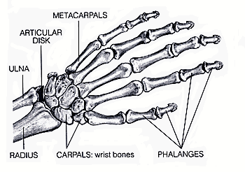
• The wrist has 8 carpal bones
• 5 metacarpal bones
• The phalanges
o The bones of the fingers and the thumb
o The Pelvic Girdle and Lower Limb
The pelvic girdle consists of 2 heavy, large coxal bones (hip bones)
The pelvis
• Is a basin composed of the pelvic girdle, sacrum, and coccyx
• Bears the weight of the body
• Protects the organs within the pelvic cavity
• Serves as the place of attachment for the legs
Each coaxl bone has 3 parts:
• The ilium
o The largest part
o Hips occur where it flares out
• The ischium
o Part that we sit on
o Has a posterior spine (ischial spine) for muscle attachment
• The pubis
o 2 pubic bones are joined together by a fibrocartilaginousjoint (the pubic symphysis)
o The male and female pelves differ from each other.
The hip socket (acetabulum) occurs where these 3 bones meet
The Femur (thighbone)
• The longest and strongest bone in the body
• The head of the femur articulates with the coxal bones at the acetabulum, and the short neck better positions the legs for walking
• Has 2 large processes
o The greater and lesser trochanters
Places of attachment for thigh muscles, buttock muscles, and hip flexors.
• At its distal end there is a medial and lateral condyles that articulate with the tibia of the leg
o This is the region of the knee and the patella (kneecap)
• The fibula
o The more slender bone in the leg
o Has a head that articulates with the tibia and the distal lateral malleous that forms the outer bulge of the ankle.
Each foot has an ankle, an instep, and five toes
• Has 7 tarsal bones
• A calcaneus (heel bone)
• Instep has 5 elongated metatarsal bones
• Bones of the toes are called phalanges.
• Articulations
o Bones are joined at the joints
Classified as:
• Fibrous
o Are immovable
• Cartilaginous
o Connected by hyaline cartilage or fibrocartilage
o Slightly movable
• Synovial
o Move freely
o Filled with synovial fluid, a lubricant for the joint
o Elbow and knee joints
o Also called hinge joints
• Chapter 12
• Overview of Muscular System

o Types of muscles
Smooth
• Muscle fibers are spindle shaped cells, with a single nucleus
• Cells are arranged in parallel lines, forming sheets
• Located in walls of hollow internal organs, it causes them to contract
• Contraction is involuntary
Cardiac
• Forms the heart wall
• Its fibers are uninucleated, striated, tubular, and branched, which allows the fibers to interlock at intercalated disks
• Fibers relax completely between contractions, which prevents fatigue.
• Contraction is rhythmical, and involuntary
Skeletal
• Fibers are tubular, multinucleated, and striated.
• Make up the skeletal muscles attached to the skeleton.
• Run the length of the muscles
• Muscle contractions are voluntary
o Functions of Skeletal Muscles
Support the body
Make bones move
Help maintain constant body temperature
Muscle contraction assists movement in cardiovascular and lymphatic vessels.
Help protect internal organs and stabilize joints
o Skeletal Muscles of the Body
Basic Structure of Skeletal Muscles
• A whole muscle contains bundles of fascicles (skeletal muscle fibers)
• Within a fascicle ach fiber is surrounded by connective tissue
• The fascicle itself is also surrounded by connective tissue
• Muscles are covered with fascia, a type of connective tissue that extends beyond the muscle and becomes its tendon
Skeletal Muscles Work in Pairs
• The origin of a muscle is on a stationary bone
• The insertion of a muscle is on a bone that moves
• When a muscle contracts, it pulls on the tendons at its insertion, and the bone moves
• Skeletal muscles function in groups
• To make a particular movement the nervous system stimulates an appropriate group of muscles
o 1 muscle does most of the work (Prime Mover)
o Other muscles, called synergists, assist the prime mover.
o When muscles contract they shorten, they can only pull, they work in opposite pairs
• Skeletal Muscle Fiber Contraction
o Muscle Fibers and How They Slide
Myofibrils and Sarcomeres
• Myofibrils
o Cylindrical in shape, and run the length of the muscle fiber
o The striations of skeletal muscle fibers are formed by the placement of myofilaments within units of myofibrils called sarcomeres
o A sarcomere extends between 2 dark lines called Z lines
o Sarcomere contains 2 types of protein myofilaments
Thick filaments
• Made of myosin
Thin filaments
• Made of actin
o The I band is light colored because it contains only actin filaments attached to a Z line. The dark regions of the A band contain overlapping actin and myosin filaments, its H zone has only myosin filaments
• Myofilaments
o Thick filaments
Composed of several hundred molecules of myosin
Each molecule is shaped like a golf club
o Thin Filaments
Consists of 2 intertwining strands of actin
2 other proteins called tropomyosin and troponin also plays a role
o Sliding Filaments
When muscles are stimulated, impulses travel down a T tubule, and calcium is released from the sarcoplasmic Reticulum.
The muscle fiber now contracts as the Sarcomere, within the myofibrils, shorten.
When a sarcomere shortens, the actin filaments slide past the myosin filaments and approach one another.
This causes the I band to shorten, the Z line to move inward, the H zone to almost or completely disappear.
During the sliding process, the sarcomere shortens, even though the filaments themselves remain the same length.
o Control of Muscle Fiber Contraction
Neuromuscular junction
• Muscle fibers are stimulated to contract by motor neurons whose axons are in nerves.
• The axon of one motor neuron can stimulate from a few to several muscle fibers of a muscle because each axon has several branches.
• Each branch of an axon ends in an axon terminal that lies in close proximity to the sarcolemma of a muscle fiber.
• The synaptic cleft separates the axon terminal from the sarcolemma.
Axon terminal contain synaptic vesicles that are filled with the neurotransmitter Acetylcholine(Ach).
When nerve impulses arrive at an axon terminal, the synaptic vesicles release Ach into the synaptic cleft.
Botox prevents wrinkling of the brow and skin about the eyes because it blocks the release of Ach into the synaptic cleft, therefore muscle contraction never occurs.
When ACh is released it diffuses across the cleft and binds to receptors in the sarcolemma.
The sarcolemma generates impulses that spread over the sarcolemma and down T tubules to the sarcoplasmic Reticulum.
The release of Ca2+ from the sarcoplasmic Reticulum leads to sarcomere contraction.
When Ca2+ ions are released from the sarcoplasmic Reticulum, they combine with troponin, this causes tropomyosin threads to shift their position, exposing myosin-binding sites.
Myosin can now bind to actin.
• Whole Muscle Contraction
o Muscles Have Motor Units
A nerve fiber, together with all of the muscle fibers it innervates, is called a motor unit.
A motor unit obeys the all-or-none law.
• All the muscle fibers in a motor unit are stimulated at once, they can either contract or they do not contract.
When a motor unit is stimulated by infrequent electrical impulses, a single contraction occurs that lasts only a fraction of a second.
• This response is called a muscle twitch.
o 3 Stages:
The Latent Period
• The period of time between stimulation and initiation of contraction
The Contraction Period
• The muscle shortens
The Relaxation Period
• The muscle returns to its former length
A whole muscle contains many motor units
As the intensity of nervous stimulation increases, more and more motor units in a muscle are activated.
o Muscle Tone
Is dependent on muscle contraction
Occurs when some motor units are always contracted but not enough to cause movement, the muscle is firm and solid
o Energy for Muscle Contraction
Fuel Sources for Exercise
• Four possible energy sources.
o Two are stored in the muscle
Glycogen and fat (triglycerides) are stored in muscles.
The intensity and duration of exercise determines which one is used.
o Two are acquired from blood
Blood glucose and plasma fatty acids are used as an energy source.
Both are delivered by circulating blood.
Sources of ATP for Muscle Contraction
• Muscle cells store limited amounts of ATP.
• There are 3 ways to acquire more ATP once the stored ATP has been used up:
o The CP (creatine phosphate) Pathway
The simplest and fastest way for muscles to produce ATP, because it consists of only one reaction.
The reaction occurs in the midst of sliding filaments.
CP is formed only when a muscle cell is resting, and only a limited amount is stored.
It is used at the beginning of submaximal exercise and during short-term, high intensity exercise that lasts less than 5 seconds.
o Fermentation
Intense activities lasting longer than 5 seconds use fermentation.
Fermentation produces 2 ATP from the breakdown of glucose to lactate anaerobically.
Is fast-acting but results in the build up of lactate, which produces short-term muscle aches and fatigue.
o Cellular Respiration
Is more likely to supply ATP when exercise is submaximal in intensity.
Makes use of glucose from the breakdown of glycogen stored in muscle, glucose taken up from the blood, and fatty acids.
o Fast-Twitch and Slow-Twitch Muscle Fibers
Fast-Twitch Fibers
• Usually anaerobic and are designed for strength because their motor fibers contain many fibers.
• They provide explosions of energy and are most helpful in sports activities.
• Light in color because they have fewer mitochondria, little or no myoglobin, and fewer blood vessels.
• Develops maximum tension more rapidly than slow-twitch fibers, maximum tension is greater.
• They fatigue quickly.
Slow-Twitch Fibers
• Have a steadier tug and have more endurance
• They produce most of their energy aerobically; they tire only when their fuel supply is gone.
• They have many mitochondria and are dark in color because they contain myoglobin.
• They are surrounded by dense capillary beds and draw more blood and oxygen than fast-twitch fibers.
• Have a low maximum tension, which develops slowly.
• Highly resistant to fatigue.
o Delayed Onset Muscle Soreness
Generally appears 24-48 hours after strenuous exercise.
Is due to tissue damage that takes several days to heal
• Muscular Disorders
o Common Muscular Conditions
Spasms
• Sudden involuntary muscular contractions often accompanied by pain.
Cramps
• Strong, painful spasms, usually due to strenuous activity.
Strain
• Stretching or tearing a muscle
Sprain
• Twisting of a joint leading to injury and swelling
Tendinitis
• The normal, smooth gliding motion of a tendon is impaired, the tendon is inflamed, movement of a joint becomes painful.
Bursitis
• Inflammation of the bursa
o Muscular Diseases
Myalgia
• Achy muscles
Fibromyalgia
• Chronic condition
• Symptoms include
o Achy pain, tenderness, stiffness of muscles
Muscular Dystrophy
• Broad term applied to a group of disorders that are characterized by a progressive degeneration and weakening of muscles
Myasthenia Gravis
• An autoimmune disease characterized by weakness that especially affects the muscles of the eyelids, face, neck, and extremities.
Amyotrophic Lateral Sclerosis
• Known as Lou Gehrig’s Disease
• Experience gradual loss of the ability to walk, talk, chew, and swallow.
• Homeostasis
o Both Systems Produce Movement
The skeletal and muscular system work together to enable body movement.
o Both Systems Protect Body Parts
The skeletal system protects the internal organs
The muscular system pads bones
o Bones Store and Release Calcium
The skeletal system performs tasks that are vital for calcium homeostasis.
o Blood Cells are Produced in Bones
Red bone marrow is the site of blood cell production.
o Muscles Help Maintain Body Temperature
When cold, smooth muscles in the blood vessels constrict, reducing amount of blood that is close to the surface of the body.
• Neurons
o Cells that transmit nerve impulses between parts of the nervous system
• Neuroglia
o Support and nourish neurons
o Neuron Structure
3 types of neurons
• Sensory neurons
o Takes nerve impulses (messages) from a sensory receptor to the CNS
Sensory receptor is a special structure that detects changes in the environment.
• Interneuron
o Is completely within the CNS, they receive input from sensory neurons and from other interneurons in the CNS.
o After receiving the information they sum it all up before translating it to motor neurons
• Motor Neuron
o Takes nerve impulses away from the CNS to an effector (muscle fiber or gland)
Effectors carry out our responses to environmental changes, external and internal.
o Myelin Sheath
Is the protective cover over the axons.
Is formed by neuroglia cells called Schwann Cells
• Schwann Cells contain myelin in their plasma membrane
• Develops when these cells wrap themselves around an axon many times.
• Each neuroglia cell only covers a part of the axon, the gaps where there is no cover is called nodes of Ranvier.
Long Axons have a myelin sheath, but short axons do not.
Myelin gives nerve fibers their white glistening appearance and is also an insulator.
The myelin sheath is important to nerve regeneration within the PNS.
• If an axon is severed, the myelin sheath remains and serves as a passageway for new fiber growth.
o The Nerve Impulse
Nerve impulses convey information within the nervous system.
• Nerve impulse is studied by using excised axons and a voltmeter to measure voltage.
o The voltmeter allows measurement of the potential difference between 2 sides of the axonal membrane
Resting potential
• Is when the axon is not conducting an impulse.
• The inside of the neuron is more negative than the outside
• Resting potential correlates to a difference in ion distribution on either side of the axonal membrane
o More Na+(sodium ions) outside the axon
o More K+ (potassium ions) inside the axon
o Sodium-Potassium Pump created the unequal distribution of these ions.
Actively transports Na+ out of the axon
Actively transports K+ into the axon
The membrane is permeable to K+ but not to Na+, causing there to always be more positive ions outside the membrane than inside.
Action Potential
• Is a rapid change in polarity across an axonal membrane as the nerve impulse occurs.
• If a stimulus causes the axonal membrane to depolarize to a certain level, called threshold, an action potential occurs in an all-or-none manner.
• Action potential requires 2 types of gated channel proteins in the membrane
o Sodium Gates Open
When action potential occurs, gates of sodium channels open first
Na+ flows into the axon.
This causes a depolarization because the charge inside the axon changes from negative to positive.
o Potassium Gates Open
Second, the gates of the potassium channels open
K+ flows to outside the axon.
This causes a repolarization because the inside of the axon resumes a negative charge as K+ exits he axon.
Visualizing an Action Potential
• After an action potential has passed by, the sodium-potassium pump restores the resting potential by moving the potassium back to the inside and sodium back to the outside.
o Propagation of an Action Potential
Action potentials are self-propagating; each action potential generates another along the length of an axon.
When an axon does not have a myelin sheath, the action potential at one locale stimulates an adjacent part of the axon’s membrane to produce an action potential.
In axons that have a myelin sheath, an action potential at one ranvier node causes an action potential at the next node.
o The Synapse
Every axon branches into many fine endings, each is tipped by a small swelling called an axon terminal.
Each axon terminal lies very close to either the dendrite or the cell body of another neuron
This region of close proximity is called a synapse
• At a synapse, a small gap called the synaptic cleft separates the sending neuron from the receiving neuron.
Transmission across a synapse is carried out by molecules called neurotransmitters.
These events occur:
• Nerve impulses traveling along an axon reach an axon terminal.
• Calcium ions enter the terminal, and they stimulate synaptic vesicles to merge with the sending membrane
• Neurotransmitter molecules are released into the synaptic cleft, and they diffuse across the cleft to the receiving membrane, where they bind with specific receptor proteins.
Neurotransmitter Molecules
• More than 100 substances are known or suspected to be neurotransmitters, some common ones are:
o Acetylcholine and norepinephrine
Active in both the CNS and PNS
Norepinephrine excites smooth muscle in the CNS
Acetylcholine in the PNS; excites skeletal muscle and inhibits cardiac muscle.
o GABA (gamma aminobutyric acid)
Is an abundant inhibitory neurotransmitter in the CNS
o Serotonin
Involved in thermoregulation, sleeping, emotions, and perception
• The Central Nervous System
o The spinal cord and the brain make up the CNS
o Sensory information is received and motor control is initiated.
o The Spinal Cord
Extends from the base of the brain through a large opening in the skull called the foramen magnum and into the vertebral canal formed by openings in the vertebrae.
Structure of the spinal cord
• Spinal nerves project from the cord between the vertebrae that make up the vertebral column.
• Central canal contains cerebrospinal fluid, along with the meninges that protect the spinal cord.
• The gray matter is centrally located and shaped like the letter H.
o This is where portions of the sensory neurons and motor neurons are found, interneurons that communicate with these 2 types of neurons are also found there.
• Dorsal root contains sensory fibers entering the gray matter
• Ventral roots contains motor fibers exiting the gray matter
• The dorsal and ventral roots join before the spinal nerve leaves the vertebral canal as a mixed nerve
• White matter occurs in areas around the gray matter
o It contains ascending tracts taking information to the brain and descending tracts taking info from the brain.
Functions of the Spinal Cord
• Provides a means of communication between the brain and the peripheral nerves that leave the cord.
• Sensory receptors generate nerve impulses that pass through sensory fibers to the spinal cord and up ascending tracts to the brain.
• Gate Control Theory of Pain:
o The tracts in the spinal cord have ”gates,” and these “gates” control the flow of pain messages from the peripheral nerves to the brain.
o Depending on how the gates process a pain signal, the pain message can be allowed to pass directly to the brain or it can be prevented from reaching the brain.
o When the brain initiates voluntarily control over our limbs, motor impulses originating in the brain pass down descending tracts to the spinal cord and out to our muscles by way of motor fibers
o If the spinal cord is severed, we suffer a loss of sensation and a loss of voluntary control.
If the cut occurs in the thoracic region, the lower body and legs are paralyzed, known as paraplegia.
If the injury is in the neck region, all four limbs are usually affected, known as quadriplegia.
o Reflex Actions
A stimulus causes sensory receptors to generate nerve impulses that travel in sensory axons to the spinal cord
Interneurons integrate the incoming data and relay signals to motor neurons
A response to the stimulus occurs when motor axons cause skeletal muscles to contract.
Each interneuron in the spinal cord has synapses with many other neurons, they send signals to several other interneurons and motor neurons.
o The Brain
The Cerebrum
• Also referred to as the Telencephalon
• Is the larges part of the brain.
• The last center to receive sensory input and carry out integration before commanding voluntary motor responses
• It communicates with and coordinates the activities of the other parts of the brain.
• Cerebral Hemispheres
o 2 halves
Left Cerebral Hemisphere
Right Cerebral Hemisphere
o A deep groove called the longitudinal fissure divides the left and right cerebral hemispheres.
o Shallow grooves called sulci divide each hemisphere into lobes
Frontal Lobe
• Is the most ventral of the lobes
• Located directly behind the forehead
Parietal lobe
• Dorsal to the frontal lobe
Occipital Lobe
• Dorsal to the parietal lobe
• Located at the real of the head
Temporal lobe
• Inferior to the front and parietal lobes
• At the temple and the ear
• The Cerebral Cortex
o Is a thin but highly convoluted outer layer of gray matter that covers the cerebral hemispheres.
o Is the region of the brain that accounts for:
Sensation
Voluntary movement
All the thought processes we associate with consciousness
• Primary Motor and Sensory Areas of the Cortex
o Primary Motor Area
Located in the frontal lobe just before the central sulcus.
Voluntary commands to skeletal muscles begin in the primary motor area, each part of the body is controlled by a certain section
o Primary Somatosensory Area
Is located dorsal to the central sulcus in the parietal lobe
Sensory information from the skin and skeletal muscles arrives here, where each part of the body is sequentially represented
• Primary taste area
o Accounts for taste sensations
• Primary visual area
o In the occipital lobe receives information from our eyes
• Primary auditory area
o In the temporal lobe receives information from our ears
• Primary olfactory area
o Is for smell, located in the temporal lobe
o Association Areas
Located in places where integration occurs
Ventral to the primary motor area is a pre-motor area
• The pre-motor area organizes motor functions for skilled motor activities, such as walking and talking at the same time
• The primary motor area then sends signals to the cerebellum, which integrates them.
o Processing Centers
Processing centers of the cortex receive information from the other association areas and perform higher-level analytical functions
The prefrontal area, an association area in the frontal lobe, receives information from other association areas and uses this information to reason and plan our actions
o Central White Matter
Most of the remaining cerebrum is composed of white matter
The Diencephalon
• Is a region that encircles the third ventricle
• Hypothalamus and the thalamus are located here
o Hypothalamus forms the floor of the third ventricle
Integrating center that regulates hunger, sleep, thirst, body temperature, and water balance,
Controls the pituitary gland, is the link between the nervous and endocrine system
o Thalamus consists of 2 masses of gray matter located in the sides and roof of the third ventricle.
Is on the receiving end for all sensory input except smell
Visual, auditory, and somatosensory information arrives at the thalamus via the cranial nerves and tracts from the spinal cord.
The thalamus integrates this information and sends it on to the appropriate portions of the cerebrum.
The Cerebellum
• Lies under the occipital lobe of the cerebrum and is separated from the brain stem by the fourth ventricle
• Made up of 2 portions that are joined by a narrow median portion
• Receives sensory input from the eyes, ears, joints, and muscles about the present position of body parts, and receives motor output from the cerebral cortex about where these parts should be located.
• After integrating this information, the cerebellum sends motor impulses by way of the brain stem to the skeletal muscles.
The Brain Stem
• Contains the midbrain, the pons, and the medulla oblongata
o Midbrain
Acts as a relay station for tracts passing between the cerebrum and the spinal cord or cerebellum.
Has reflex centers for visual, auditory, and tactile responses
o Pons
Contains bundles of axons traveling between the cerebellum and the rest of the CNS.
Functions with the medulla oblongata to regulate breathing rate and has reflex centers concerned with head movements in response to visual and auditory stimuli
o Medulla Oblongata
Contains a number of reflex centers for regulating heartbeat, breathing, and blood pressure.
Contains reflex centers for vomiting, coughing, sneezing, hiccupping, and swallowing.
• The Limbic System and Higher Mental Functions
o Limbic System
An evolutionary ancient group of linked structures deep within the cerebrum that is a functional grouping rather than an anatomical one
It blends primitive emotions and higher mental functions into a united whole.
There are two structures within the limbic system
• The amygdala
o Can cause experiences to have emotional overtones
o Creates the sensation of fear.
• The Hippocampus
o Believed to play a crucial role in learning and memory
Acts as an information gateway during the learning process, determining what information about the world is to be sent to memory, and how the information is to be encoded and stored by other regions in the brain
o Higher Mental Functions
Types of memory
• Short-term memory
• Long-term memory
• Semantic memory: number, words
• Episodic memory: persons, events
• Skill Memory: involved in performing motor activities
Long-Term Memory Storage and Retrieval
• Long-term memories are apparently stored in bits and pieces throughout the sensory association areas of the cerebral cortex.
• The Peripheral Nervous System
o Nerves that lie outside the CNS
o 2 types
Cranial Nerves
• Attached to the brain
• 12 pairs
Spinal Nerves
• Emerge in 31 pairs from either side of the spinal cord.
o Somatic System
Nerves in the somatic system serve the skin, skeletal muscles, and tendons.
Includes nerves that take sensory information from external sensory receptors to the CNS and motor commands away from the CNS to the skeletal muscles.
Automatic responses to a stimulus in the somatic system are called reflexes
• The Reflex Arc
o The path of a reflex that involves only the spinal cord.
o Sensory receptors in the skin generate nerve impulses that move along sensory fibers through the dorsal-root ganglia toward the spinal cord.
o Sensory neurons that enter the cord dorsally pass signals on to many interneurons.
o Nerves impulses travel along motor fibers to an effector, which brings about a response to the stimulus.
o The whole series of responses occurs because some of the interneurons involved carry nerve impulses to the brain.
o Autonomic System
Located in the PNS
Regulates the activity of cardiac and smooth muscles and glands
Divided into the sympathetic and parasympathetic divisions.
• Shared features are:
o They function automatically and in an involuntary manner
o They innervate all internal organs
o They utilize 2 neurons and one ganglion for each impulse
• Sympathetic division
o Important to fight or flight situations
o Accelerates the heart beat and dilates the bronchi
• Parasympathetic Division
o Promotes all the internal responses we associate with a relaxed state
• Chapter 14
• Sensory Receptors and Sensations
o Sensory Receptors
Are dendrites that are specialized to detect certain types of stimuli
Exteroceptors
• Sensory receptors that detect stimuli from outside the body
• Ex: those that result in taste, smell, vision, hearing, and equilibrium
• Are not directly involved in homeostasis and continually send messages to the central nervous system regarding environmental conditions
Interoceptors
• Receive stimuli from inside the body
• Ex: pressoreceptors respond to changes in blood pressure, osmoreceptorsdetect changes in water-balance
• Are directly involved in homeostasis and are regulated by a negative feedback mechanism
o Types of Sensory Receptors
Chemoreceptors
• Respond to chemical substances in the immediate vicinity
• Ex: taste, smell,
• Pain Receptors
o Are a type of chemoreceptor, they are naked dendrites that respond to chemicals released by damaged tissues
Photoreceptors
• Respond to light energy
• Our eyes contain photoreceptors that are sensitive to light rays and provide us with a sense of vision.
Mechanoreceptors
• Are stimulated by mechanical forces, which most often result in pressure of some sort.
Thermoreceptors
• Are located in the hypothalamus and skin and are stimulated by changes in temperature.
o Warmth receptors respond when temperatures rise
o Cold receptors respond when temperatures lower
o How Sensation Occurs
Sensory receptors respond to environmental stimuli by generating nerve impulses.
When nerve impulses arrive at the cerebral cortex of the brain, sensation, which is the conscious perception of stimuli, occurs.
All sensory receptors initiate nerve impulses; the sensation that results depends on the part of the brain receiving the nerve impulses
• Proprioceptors and Cutaneous Receptors
o Proprioceptors
Are mechanoreceptors involved in reflex actions that maintain muscle tone, and thereby the body’s equilibrium and posture.
They help us know the position of our limbs in space by detecting the degree of muscle relaxation, the stretch of tendons, and the movement of ligaments.
Muscle spindles increase the degree of muscle contraction
Golgi tendon organs decrease the degree of muscle contraction
o Cutaneous Receptors
Are located in the dermis, making the skin sensitive to touch, pressure, pain, and temperature.
There are 3 types of receptors that are sensitive to fine touch
• Meissner corpuscles
o Along with Krause end bulbs are concentrated in the fingertips, the palms, the lips, the tongue, the nipples, the penis, and the clitoris.
• Merkel disks
o Located where the epidermis meets the dermis
• Root Hair Plexus
o Is a free nerve ending that winds around the base of a hair follicle and fires if the hair is touched.
2 types of receptors that are sensitive to pressure
• Pacinian corpuscles
o Are onion shaped receptors that lie deep inside the dermis.
• Ruffini Endings
o Are encapsulated by sheaths of connective tissue and contain lacy networks of nerve fibers.
o Pain Receptors
Nociceptors
• Pain receptors for internal organs that are sensitive to chemicals released by damaged tissues.
• When inflammation occurs, because of mechanical, thermal, or electrical stimuli or toxic substances, cells release chemicals that stimulate pain receptors.
• Senses of Taste and Smell
o Taste and smell are chemical senses because their receptors are sensitive to molecules in the food we eat and the air we breathe.
o Chemoreceptors are plasma membrane receptors that bind to particular molecules. They are divided into 2 types:
Those that respond to distant stimuli
• Olfactory cells act from a distance
Those that respond to direct stimuli
• Taste cells act directly
o Sense of Taste
Taste buds are primarily located on the tongue
Four Primary types of taste
• Sweet
• Sour
• Salty
• Bitter
• A fifth taste called umami may exist for certain flavors of:
o Cheese
o Beef broth
o Some sea food
Certain regions of the tongue are more sensitive to different types of taste.
How the Brain Receives Taste Information
• Taste buds open at a taste pore.
• They have supporting cells and a number of elongated taste cells that end in microvilli.
• When molecules bind to receptor proteins of microvilli nerve impulses are generated in sensory nerve fibers that go to the brain.
• When they reach the gustatory (taste) cortex, they are interpreted as particular tastes.
o Sense of Smell
80-90% of what we perceive as taste is actually due to the sense of smell.
Our sense of smell depends on 10-20 million olfactory cells located within olfactory epithelium high in the roof of the nasal cavity.
How the Brain receives Odor Information
• Each olfactory cell has one out of several hundred different types of receptor proteins.
• Nerve fibers from like olfactory cells lead to the same neuron in the olfactory bulb, an extension of the brain.
• An odor contains many odor molecules, which activate a characteristic combination of receptor proteins.
• An odor’s signature in the olfactory bulb is determined by which neurons are stimulated.
• When neurons communicate this information via the olfactory tract to the olfactory areas of the cerebral cortex.
• Sense of Vision
o Anatomy and Physiology of the Eye
Is an elongated sphere about 2.5 cm in diameter
Has 3 Layers
• The Sclera
o The outer layer
o Is white and fibrous
o Except for the cornea, which is made of transparent collagen fibers
• The Choroid
o The middle, thin darkly pigmented layer
o Is vascular and absorbs stray light rays that photoreceptors have not absorbed.
o Toward the front it becomes the iris.
Regulates the size of the pupil
Pupil is a hole in the center of the iris that lets light into the eyeball.
o Behind the iris the choroids thickens and becomes ciliary body
Controls the shape of the lens for near and far vision
o The lens, divides the eye into 2 compartments
The anterior compartment
• Is filled with a clear watery fluid called aqueous humor
• Located in front of the lens
Posterior Compartment
• Located behind the lens
• Is filled with vitreous humor, a clear gelatinous material
• The Retina
o Located in the posterior compartment
o Contains photoreceptors called
Rod Cell
• Very sensitive to light
• Do not see color
Cone Cells
• Require bright light
• Sensitive to different wavelengths of light, giving us the ability to distinguish colors.
o Fovea centralis
Region where cone cells are packed densely
Light is normally focused here when we directly look at an object
o Optic Nerve
Formed from sensory fibers in the retina
Takes nerve impulses to the visual cortex
Function of the Lens
• The cornea, assisted by the lens and humors, focuses images on the retina.
• Visual Accommodation
o Occurs for close vision
o The lens rounds up, in order to bring the image to focus on the retina.
o The ciliary muscle contracts, releasing tension on the suspensory ligaments, the lens rounds up due to the natural elasticity.
• Sense of Hearing
o The ear has 2 sensory functions:
Hearing
Balance
The sensory receptors for both are located in the inner ear
Each consists of hair cells with stereocilia that are sensitive to mechanical stimulation. They are mechanoreceptors
o Anatomy and Physiology of the Ear
The ear has 3 divisions
• Outer
o Outer ear consists of the pinna and the auditory canal
• Middle
o Begins at the tympanic membrane and ends at a bony wall containing two small openings covered by membranes
These openings are called the:
• Oval window
• Round window
o Three small bones are found between the tympanic membrane and the oval window. They are called the ossicles
Malleus (hammer)
Incus (anvil)
Stapes (stirrup)
o The Eustachian Tube is an auditory tube that extends from the middle ear to the nasopharynx, it permits equalization of air pressure
• Inner
o Is filled with fluid
o Has three areas:
Semicircular canals
• Concerned with equilibrium
Vestibule
• Concerned with equilibrium
Cochlea
• Concerned with hearing
o Auditory Pathway to the Brain
Through the Auditory Canal and Middle Ear
• The process begins when sound waves enter the auditory canal.
• When a large number of waves strike the tympanic membrane, it moves back and forth ever so slightly.
• The malleus then takes the pressure from the inner surface of the tympanic membrane and passes it to the stapes.
• The stapes strikes the membrane of the oval window, causing it to vibrate; the pressure is passed to the fluid within the cochlea.
From the Cochlea to the Auditory Cortex.
• Cochlea has 3 canals
o The cochlear canal
o The vestibular canal
o The tympanic canal
• When the stapes strikes the membrane of the oval window, pressure waves move from the vestibular canal to the tympanic canal across the basilar membrane.
• The basilar membrane moves up and down, and the stereocilia of the hair cells embedded in the tectorial membrane bend.
• Nerve impulses begin in the cochlear nerve and travel to the brain.
• When they reach the auditory cortex in the temporal lobe, they are interpreted as a sound.
• Sense of Equilibrium
o Rotational Equilibrium Pathway
Mechanoreceptors in the semicircular canals detect rotational and angular movement of the head.
The three semicircular canals are arranged so that there is one in each dimension of space
The base of each of the three canals, called the ampulla, is slightly enlarged.
Little hair cells, whose stereocilia are embedded within a gelatinous material cupula, are found within the ampullae.
Because of the way the semicircular canals are arranged, each ampulla responds to head rotation in a different plane of space.
As fluid within a semicircular canal flows over and displaces a cupula, the stereocilia of the hair cells bend, and the pattern of impulses carried by the vestibular nerve to the brain changes.
The brain uses information from the hair cells within ampulla of the semicircular canals to maintain equilibrium through appropriate motor output to various skeletal muscles that can right our present position in space as need be.
o Gravitational Equilibrium Pathway
The mechanoreceptors in the utricle and saccules detect movement of the head in the vertical or horizontal planes.
The utricle and saccules are two membraneous sacs located in the inner ear near the semicircular canals.
Both of these sacs contain little hair cells, whose stereocilia are embedded within a gelatinous material called an otolithic membrane. Calcium carbonate granules, otoliths, rest on this membrane.
When the body is still, the otoliths in the utricle and the saccules rest on the otolithic membrane above the hair cells
When the head bends or the body moves in the horizontal and vertical planes, the otoliths are displaced and the otolithic membrane sags, bending the stereocilia of the hair cells beneath.
If the stereocilia move toward largest stereocilium, called the kinocilium, nerve impulses increase in the vestibular nerve.
If the stereocilia move away from the kinocilium, nerve impulses decrease in the vestibular nerve.
The frequency of nerve impulses in the vestibular nerve indicates whether you are moving up or down.
These data reach the brain, which uses them to determine the direction of the movement of the head at the moment.
The brain uses this information to maintain gravitational equilibrium through appropriate motor output to various skeletal muscles that can right our present position in space as need be.
• Chapter 11
• Overview of Skeletal System
o Functions of the Skeleton
The skeleton supports the body.
• Bones of the legs support the entire body when we are standing.
• The bones of the pelvic girdle support the abdominal cavity.
The skeleton protects soft body parts.
• Bones of the skull protect the brain
• The rib cage protects the heart and lungs.
• The vertebrae protect the spinal cord.
The skeleton produces blood cells
• All bones in the fetus have red bone marrow that produces blood cells
• Only certain bones in adults produce blood cells.
The skeleton stores mineral and fat
The skeleton, along with the muscles, permits flexible body movement.
o Anatomy of a Long Bone
The diaphysis
• The shaft, or main portion of the bone
• Has a large medullary cavity
o Walls are composed of compact bone
o Lined with a thin, vascular membrane, and is filled with yellow bone marrow that stores fat.
The epiphyses
• Expanded region at the end of a long bone
• Composed largely of spongy bone that contains red bone marrow
• Coated with a thin layer of hyaline cartilage, called articular cartilage
Periosteum
• A layer of fibrous connective tissue covering the long bone
• Contains blood vessels, lymphatic vessels, and nerves
• Is continuous with ligaments and tendons that are connected to a bone.
Bone
• Compact Bone
o Highly organized and composed of tubular units called osteons.
• Spongy Bone
o Unorganized appearance
o It contains numerous thin plates called trabeculae, separated by unequal spaces
Cartilage
• Not as strong as bone
• More flexible than bones
• Has no nerves
• Made to pad joints where the stresses of movement are intense.
• Three types of cartilage
o Hyaline Cartilage
Firm and somewhat flexible
The matrix is uniform and glassy
Found at the ends of long bones, in the nose, at the end of the ribs, and in the larynx and trachea.
o Fibrocartilage
Stronger than hyaline
Able to withstand tension and pressure
Found in the disks located between the vertebrae and in the cartilage of the knee.
o Elastic cartilage
More flexible than hyaline cartilage
Found in the ear flaps and the epiglottis
• Fibrous connective tissue
o Contains rows of cells called fibroblasts separated by bundles of collagenous fibers
o Makes up the ligaments that connect bone to bone.
o Makes up the tendons that connect muscles to a bone at joints (articulations).
• Bone Growth, Remodeling, and Repair
o Several different types of bone cells are involved in bone growth, remodeling, and repair.
Osteoblasts
• Bone-forming cells
• They secrete the organic matrix of bone and promote the deposition of calcium salts into the matrix.
Osteocytes
• Mature bone cells
• Derived from osteoblasts
• Maintain the structure of bone
Osteoclasts
• Bone-absorbing cells
• They break down bone and assist in depositing calcium and phosphate in the blood.
o Bone Growth and Development
Ossification
• The formation of bone.
The bones of the skeleton form during embryonic development in 2 ways:
• Intramembranous Ossification
o Flat bones are examples of intramembranous Ossification.
o Bones develop between sheets of fibrous connective tissue.
o Cells derived from connective tissue cells become osteoblasts located in the ossification centers.
o Osteoblasts secrete the organic matrix of bone consisting of mucopolysaccharides and collagen fibrils.
o Calcification occurs when calcium salts are added to the organic matrix
o Osteoblasts promote calcification
o Ossification results in the trabeculae of spongy bone, spongy bone remains on the inside.
o Periosteum forms outside the spongy bone and osteoblasts, derived from the periosteum, carry out further ossification.
o Trabeculae form and fuse to become compact bone, which surrounds the spongy bone.
• Endochondral Ossification
o How most of the bones of the human skeleton are formed.
o Bone replaces cartilaginous models of the bones.
o Gradually, the cartilage is replaced by the calcified bone matrix that makes these bones capable of bearing weight.
• Epiphyseal Plates
o Have four layers
Resting Zone
• Layer nearest the epiphysis
• Where cartilage remains
Proliferating Zone
• Next to the resting zone
• Chondrocytes are producing new cartilage cells.
Degenerating Zone
• The third layer
• The cartilage cells are dying off
Ossification Zone
• Fourth layer
• Bone is forming
• Final Size of the Bones
o When epiphyseal plates close, bone length can no longer occur.
o The arms and legs of women close at about age 18.
o The arms and legs of men close at about age 20.
o Portions of other types of bones may continue until age 25.
o Hormones control the activity of the epiphyseal plate.
• Hormones Affect Bone Growth
o Several different hormones are involved in bone growth.
Growth Hormone (GH)
• Directly stimulates growth of the epiphyseal plate, and bone growth in general.
Thyroid Hormone
• GH would be ineffective if the metabolic activity of cells is not promoted.
• Thyroid Hormone promotes the metabolic activity of cells.
o Bone Remodeling and Its Role in Homeostasis
Bone Remodeling
• The process of bone renewal to keep bones strong.
• Allows the body to regulate the amount of calcium in the blood.
• The blood calcium level is critical because:
o If calcium concentration is too high, neurons and muscle cells no longer function
o If calcium concentrations fall too low, convulsions occur.
• Bones absorb excess calcium if level is too high.
• Bones release calcium if level is too low.
o Bone Repair
Is required after bone breaks or is fractured
Fracture repair takes several months in a series of 4 steps:
• Hematoma
o After a fracture blood escapes from ruptured blood vessels and forms a hematoma in the space between the ends of the broken bone
o Happens within 6-8 hours.
• Fibrocartilaginous Callus
o Tissue repair begins
o Fibrocartilaginous callus fills the space between the ends of the broken bone for about 3 weeks.
• Bony Callus
o Osteoblasts produce trabeculae of spongy bone and convert the Fibrocartilage callus to a bony callus that joins the broken bones together.
o Lasts about 3-4 months.
• Remodeling
o Osteoblasts build new compact bone at the periphery, and osteoclasts absorb the spongy bone, creating a new medullary cavity.
• Bones of the Axial Skeleton
o Lies in the midline of the body.
o Consists of the skull, hyoid bone, vertebral column, and the rib cage.
o The Skull
Formed by the cranium (braincase) and the facial bones.
The Cranium
• Protects the brain
• Composed of eight bones
o Frontal bone
Forms the forehead
o Parietal bones
Extend to the sides
o Occipital bone
Forms the base of the skull
o Temporal bone
Has an opening that leads to the middle ear
o Sphenoid bone
Extends across the floor of the cranium
Completes the sides of the skull
Contributes to forming the eye sockets
o Ethmoid bone
Lies in front of the sphenoid
Helps from the orbits and the nasal septum
The Facial Bones
• Most prominent of the facial bones are:
o Mandible
Lower jaw
Only movable part of the skull
o The maxillae
Forms the upper jaw and the anterior portion of the hard palate
o Palatine bones
Make up the posterior portion of the hard palate and the floor of the nose.
o Zygomatic bones
Cheekbone prominences
o Nasal bones
Form the bridge of the nose
The Hyoid Bone
• Only bone in the body that does not articulate with another bone.
• It is attached to the temporal bones by muscles and ligaments and to the larynx by a membrane
• Anchors the tongue and serves as the site for the attachment of muscles associated with swallowing.
The Vertebral Column
• Consists of 33 vertebrae
• Have 4 curvatures that provide more resilience and strength for an upright posture.
• Types of vertebrae
o Cervical vertebrae
Located in the neck
First cervical vertebrae, called atlas, hold up the head.
Second cervical vertebrae, called axis, allows a degree of rotation
o Thoracic Vertebrae
Have long, thin, spinous processes and articular facets for the attachment of the ribs.
o Lumbar Vertebrae
o Sacral Vertebrae
Five vertebrae fused together
o Coccyx (tailbone)
Composed of four fused vertebrae
• Intervertebral Disks
o Located between the vertebrae
o Composed of fibrocartilage that acts as a kind of padding.
o They prevent the vertebrae from grinding against each other.
o They absorb shock caused by movements.
o Allows the vertebrae to move.
The Rib Cage
• Also called the thoracic cage
• Composed of thoracic vertebrae, the ribs, and associated cartilages, and the sternum.
• Protects the heart and lungs
• The Ribs
o Flattened bone
o Originates at the thoracic vertebrae and proceeds toward the anterior thoracic wall.
o 12 pairs of ribs
Connected directly to the thoracic vertebrae in the back.
Upper 7 pairs connect to the sternum
• Referred to as the “true ribs”
Next 3 ribs connect to the sternum by a common cartilage
• Referred to as the “false ribs”
Last 2 pairs, are not attached to the sternum, they are attached to T12, the thoracic vertebrae
• Referred to as the “floating ribs”
• The Sternum
o Lies in the midline of the body
o Helps protect the heart and lungs
o Is a flat bone in the shape of a knife
o Composed of three bones
Manubrium
• The handle
The body
• The blade
Xiphoid process
• The point of the blade
• Bones of the Appendicular Skeleton
o Composed of the bones within the pectoral and pelvic girdles and their attached limbs
Pectoral girdle and upper limb are specialized for flexibility
The pelvic girdle and lower limbs are specialized for strength
o The Pectoral Girdle and Upper Limb
The body has a left and right pectoral girdles
• Each consists of:
o A scapula (shoulder blade)
o Clavicle (collarbone)
Extends across the top of the thorax
It articulates with the sternum and the acromion process of the scapula.
The muscles of the arm and chest attach to the coracoid process of the scapula.
The glenoid cavity of the scapula articulates with the head of the humorous.
• This allows the arm to move in almost any direction, but reduces stability.
Components of the pectoral girdle
• Humorous
o The single long bone in the arm
o Has a smooth round head that fits into the glenoid cavity of the scapula
o On the shaft is a tuberosity that attaches the deltoid muscle
o The far end has 2 protuberances where the radius and ulna attach
• Radius
• Ulna
The hand
• The wrist has 8 carpal bones
• 5 metacarpal bones
• The phalanges
o The bones of the fingers and the thumb
o The Pelvic Girdle and Lower Limb
The pelvic girdle consists of 2 heavy, large coxal bones (hip bones)
The pelvis
• Is a basin composed of the pelvic girdle, sacrum, and coccyx
• Bears the weight of the body
• Protects the organs within the pelvic cavity
• Serves as the place of attachment for the legs
Each coaxl bone has 3 parts:
• The ilium
o The largest part
o Hips occur where it flares out
• The ischium
o Part that we sit on
o Has a posterior spine (ischial spine) for muscle attachment
• The pubis
o 2 pubic bones are joined together by a fibrocartilaginousjoint (the pubic symphysis)
o The male and female pelves differ from each other.
The hip socket (acetabulum) occurs where these 3 bones meet
The Femur (thighbone)
• The longest and strongest bone in the body
• The head of the femur articulates with the coxal bones at the acetabulum, and the short neck better positions the legs for walking
• Has 2 large processes
o The greater and lesser trochanters
Places of attachment for thigh muscles, buttock muscles, and hip flexors.
• At its distal end there is a medial and lateral condyles that articulate with the tibia of the leg
o This is the region of the knee and the patella (kneecap)
• The fibula
o The more slender bone in the leg
o Has a head that articulates with the tibia and the distal lateral malleous that forms the outer bulge of the ankle.
Each foot has an ankle, an instep, and five toes
• Has 7 tarsal bones
• A calcaneus (heel bone)
• Instep has 5 elongated metatarsal bones
• Bones of the toes are called phalanges.
• Articulations
o Bones are joined at the joints
Classified as:
• Fibrous
o Are immovable
• Cartilaginous
o Connected by hyaline cartilage or fibrocartilage
o Slightly movable
• Synovial
o Move freely
o Filled with synovial fluid, a lubricant for the joint
o Elbow and knee joints
o Also called hinge joints
• Chapter 12
• Overview of Muscular System
o Types of muscles
Smooth
• Muscle fibers are spindle shaped cells, with a single nucleus
• Cells are arranged in parallel lines, forming sheets
• Located in walls of hollow internal organs, it causes them to contract
• Contraction is involuntary
Cardiac
• Forms the heart wall
• Its fibers are uninucleated, striated, tubular, and branched, which allows the fibers to interlock at intercalated disks
• Fibers relax completely between contractions, which prevents fatigue.
• Contraction is rhythmical, and involuntary
Skeletal
• Fibers are tubular, multinucleated, and striated.
• Make up the skeletal muscles attached to the skeleton.
• Run the length of the muscles
• Muscle contractions are voluntary
o Functions of Skeletal Muscles
Support the body
Make bones move
Help maintain constant body temperature
Muscle contraction assists movement in cardiovascular and lymphatic vessels.
Help protect internal organs and stabilize joints
o Skeletal Muscles of the Body
Basic Structure of Skeletal Muscles
• A whole muscle contains bundles of fascicles (skeletal muscle fibers)
• Within a fascicle ach fiber is surrounded by connective tissue
• The fascicle itself is also surrounded by connective tissue
• Muscles are covered with fascia, a type of connective tissue that extends beyond the muscle and becomes its tendon
Skeletal Muscles Work in Pairs
• The origin of a muscle is on a stationary bone
• The insertion of a muscle is on a bone that moves
• When a muscle contracts, it pulls on the tendons at its insertion, and the bone moves
• Skeletal muscles function in groups
• To make a particular movement the nervous system stimulates an appropriate group of muscles
o 1 muscle does most of the work (Prime Mover)
o Other muscles, called synergists, assist the prime mover.
o When muscles contract they shorten, they can only pull, they work in opposite pairs
• Skeletal Muscle Fiber Contraction
o Muscle Fibers and How They Slide
Myofibrils and Sarcomeres
• Myofibrils
o Cylindrical in shape, and run the length of the muscle fiber
o The striations of skeletal muscle fibers are formed by the placement of myofilaments within units of myofibrils called sarcomeres
o A sarcomere extends between 2 dark lines called Z lines
o Sarcomere contains 2 types of protein myofilaments
Thick filaments
• Made of myosin
Thin filaments
• Made of actin
o The I band is light colored because it contains only actin filaments attached to a Z line. The dark regions of the A band contain overlapping actin and myosin filaments, its H zone has only myosin filaments
• Myofilaments
o Thick filaments
Composed of several hundred molecules of myosin
Each molecule is shaped like a golf club
o Thin Filaments
Consists of 2 intertwining strands of actin
2 other proteins called tropomyosin and troponin also plays a role
o Sliding Filaments
When muscles are stimulated, impulses travel down a T tubule, and calcium is released from the sarcoplasmic Reticulum.
The muscle fiber now contracts as the Sarcomere, within the myofibrils, shorten.
When a sarcomere shortens, the actin filaments slide past the myosin filaments and approach one another.
This causes the I band to shorten, the Z line to move inward, the H zone to almost or completely disappear.
During the sliding process, the sarcomere shortens, even though the filaments themselves remain the same length.
o Control of Muscle Fiber Contraction
Neuromuscular junction
• Muscle fibers are stimulated to contract by motor neurons whose axons are in nerves.
• The axon of one motor neuron can stimulate from a few to several muscle fibers of a muscle because each axon has several branches.
• Each branch of an axon ends in an axon terminal that lies in close proximity to the sarcolemma of a muscle fiber.
• The synaptic cleft separates the axon terminal from the sarcolemma.
Axon terminal contain synaptic vesicles that are filled with the neurotransmitter Acetylcholine(Ach).
When nerve impulses arrive at an axon terminal, the synaptic vesicles release Ach into the synaptic cleft.
Botox prevents wrinkling of the brow and skin about the eyes because it blocks the release of Ach into the synaptic cleft, therefore muscle contraction never occurs.
When ACh is released it diffuses across the cleft and binds to receptors in the sarcolemma.
The sarcolemma generates impulses that spread over the sarcolemma and down T tubules to the sarcoplasmic Reticulum.
The release of Ca2+ from the sarcoplasmic Reticulum leads to sarcomere contraction.
When Ca2+ ions are released from the sarcoplasmic Reticulum, they combine with troponin, this causes tropomyosin threads to shift their position, exposing myosin-binding sites.
Myosin can now bind to actin.
• Whole Muscle Contraction
o Muscles Have Motor Units
A nerve fiber, together with all of the muscle fibers it innervates, is called a motor unit.
A motor unit obeys the all-or-none law.
• All the muscle fibers in a motor unit are stimulated at once, they can either contract or they do not contract.
When a motor unit is stimulated by infrequent electrical impulses, a single contraction occurs that lasts only a fraction of a second.
• This response is called a muscle twitch.
o 3 Stages:
The Latent Period
• The period of time between stimulation and initiation of contraction
The Contraction Period
• The muscle shortens
The Relaxation Period
• The muscle returns to its former length
A whole muscle contains many motor units
As the intensity of nervous stimulation increases, more and more motor units in a muscle are activated.
o Muscle Tone
Is dependent on muscle contraction
Occurs when some motor units are always contracted but not enough to cause movement, the muscle is firm and solid
o Energy for Muscle Contraction
Fuel Sources for Exercise
• Four possible energy sources.
o Two are stored in the muscle
Glycogen and fat (triglycerides) are stored in muscles.
The intensity and duration of exercise determines which one is used.
o Two are acquired from blood
Blood glucose and plasma fatty acids are used as an energy source.
Both are delivered by circulating blood.
Sources of ATP for Muscle Contraction
• Muscle cells store limited amounts of ATP.
• There are 3 ways to acquire more ATP once the stored ATP has been used up:
o The CP (creatine phosphate) Pathway
The simplest and fastest way for muscles to produce ATP, because it consists of only one reaction.
The reaction occurs in the midst of sliding filaments.
CP is formed only when a muscle cell is resting, and only a limited amount is stored.
It is used at the beginning of submaximal exercise and during short-term, high intensity exercise that lasts less than 5 seconds.
o Fermentation
Intense activities lasting longer than 5 seconds use fermentation.
Fermentation produces 2 ATP from the breakdown of glucose to lactate anaerobically.
Is fast-acting but results in the build up of lactate, which produces short-term muscle aches and fatigue.
o Cellular Respiration
Is more likely to supply ATP when exercise is submaximal in intensity.
Makes use of glucose from the breakdown of glycogen stored in muscle, glucose taken up from the blood, and fatty acids.
o Fast-Twitch and Slow-Twitch Muscle Fibers
Fast-Twitch Fibers
• Usually anaerobic and are designed for strength because their motor fibers contain many fibers.
• They provide explosions of energy and are most helpful in sports activities.
• Light in color because they have fewer mitochondria, little or no myoglobin, and fewer blood vessels.
• Develops maximum tension more rapidly than slow-twitch fibers, maximum tension is greater.
• They fatigue quickly.
Slow-Twitch Fibers
• Have a steadier tug and have more endurance
• They produce most of their energy aerobically; they tire only when their fuel supply is gone.
• They have many mitochondria and are dark in color because they contain myoglobin.
• They are surrounded by dense capillary beds and draw more blood and oxygen than fast-twitch fibers.
• Have a low maximum tension, which develops slowly.
• Highly resistant to fatigue.
o Delayed Onset Muscle Soreness
Generally appears 24-48 hours after strenuous exercise.
Is due to tissue damage that takes several days to heal
• Muscular Disorders
o Common Muscular Conditions
Spasms
• Sudden involuntary muscular contractions often accompanied by pain.
Cramps
• Strong, painful spasms, usually due to strenuous activity.
Strain
• Stretching or tearing a muscle
Sprain
• Twisting of a joint leading to injury and swelling
Tendinitis
• The normal, smooth gliding motion of a tendon is impaired, the tendon is inflamed, movement of a joint becomes painful.
Bursitis
• Inflammation of the bursa
o Muscular Diseases
Myalgia
• Achy muscles
Fibromyalgia
• Chronic condition
• Symptoms include
o Achy pain, tenderness, stiffness of muscles
Muscular Dystrophy
• Broad term applied to a group of disorders that are characterized by a progressive degeneration and weakening of muscles
Myasthenia Gravis
• An autoimmune disease characterized by weakness that especially affects the muscles of the eyelids, face, neck, and extremities.
Amyotrophic Lateral Sclerosis
• Known as Lou Gehrig’s Disease
• Experience gradual loss of the ability to walk, talk, chew, and swallow.
• Homeostasis
o Both Systems Produce Movement
The skeletal and muscular system work together to enable body movement.
o Both Systems Protect Body Parts
The skeletal system protects the internal organs
The muscular system pads bones
o Bones Store and Release Calcium
The skeletal system performs tasks that are vital for calcium homeostasis.
o Blood Cells are Produced in Bones
Red bone marrow is the site of blood cell production.
o Muscles Help Maintain Body Temperature
When cold, smooth muscles in the blood vessels constrict, reducing amount of blood that is close to the surface of the body.
Monday, March 24, 2008
Unit 2 lab project
My base line pulse was 92 bpm and my respirations were 14 per minute.
The First activity that I did was 10 situps, the second activity I did was 10 jumping jacks, and the third activity that I did was 10 push-ups.
How Do I think my ,etabolic rates will compare to my baseline rate after:

Here is a graph showing the results from the actvities

Project Analysis
For the first activity I hypothesiized that my pulse would be at 94bpm and my respirations would be at 18. I was correct for the pulse but I was one off for respirations, they were slower than I hypothesized.
For the second activity I hypothesized that my pulse would be 98/bpm and respirations would be 20. They were quite a bit lower for both pulse and respirations.
For the third activity I hypothesized that my pulse would be 93bpm and my respirations would be 17. I was correct about the pulse but the respirations were higher.
This exercise tells me that 10 sit-ups and 10 push-ups cause you to exert about the same amount of energy, jumping jacks took the least amount of energy.
Table of contents
•Cardiovascular system : heart and blood vessels
oOverview
Circulation
Functions of the cardiovascular system
Lymphatic system
oTypes of blood vessels
The arteries from the heart
The capillaries
Veins to the heart
oThe heart
Hearts anatomy
Passage of blood through the heart
oThe heart beat
Internal control
External control
oFeatures of the cardiovascular system
Pulse rate
Regulation of blood flow
Blood flow is slow in capillaries
Blood flow in veins returns blood to the heart
oCardiovascular pathways
Pulmonary circuit
•Path of blood through the lungs
•Systemic circuit
oExchange at the capillaries
oCardiovascular disorders
Disorders of the blood vessels
Disorders of the heart
•Blood
oOverview
Functions of blood
Composition of blood
oRed Blood Cells and transport of oxygen
How RBC’s carry oxygen
How RBC’s help transport carbon dioxide
RBC’s are produced in the bone marrow
Disorders involving RBC’s
oWhite Blood Cells and defense against disease
White Blood Cells (WBC)
Types of WBC’s
Disorders involving WBC’s
oPlatelets and blood clotting
Platelets
Blood clotting
Disorders relating to blood clotting
oBlood typing and transfusion
Transfusions
Blood typing
•Lymphatic system and immunity
oMicrobes, pathogens, and the body
Microbes
Pathogens
Bacteria
Viruses
Prions
oLymphatic system
oLymphatic vessels
oLymphatic organs
Primary organs
Secondary organs
oNonspecific defenses
Immunity
Inflammatory response
oSpecific defenses
How they work
B-Cells and antibody-mediated immunity
T-Cells and Cell Mediated immunity
•Cloning of T Cells
•Cytotoxic T Cells
•Helper T Cells
oAcquired immunity
Active immunity
Passive immunity
•Digestive system and nutrition
oOverview
5 processes needed for the digestive process
Wall of the digestive tract
o1st part of the digestive tract
Mouth
Pharynx and esophagus
Stomach and small intestines
•Stomach
•Small intestines
Three accessory organs and regulation of secretions
Pancreas
Liver
Gallbladder
Regulation of digestive secretions
oLarge intestines and defacation
Large intestine
Disorders of the colon and rectum
oNutrition and weight control
How obesity is defined
Classes of nutrients
•Carbohydrates
Proteins
•Lipids
•Minerals
•Vitamins
How to plan a nutritious meal
• Cardiovascular System: Heart and Blood Vessels
o Cardio vascular system consists of:
Heart: pumps the blood
Blood vessels: what the blood travels through.
o Overview
Circulation performs exchanges
• Blood exchanges substances with tissue fluid, not directly with cells.
• Blood removes waste products from the tissue fluid.
• Blood brings tissue fluid oxygen and nutrients that the cells need to continue living.
• Blood is refreshed at the lungs, intestines, and kidneys
o Lungs
Carbon dioxide leaves blood and oxygen enters.
o Kidneys
Blood is purified of its wastes; water and salt is retaind as needed.
o Intestines
Nutrients enter the blood.
• The liver
o takes amino acids from the blood and returns proteins to the blood.
o Removes poisones that entered the blood in the intestines.
Function of the cardiovascular System
• Contractions of the heart create blood pressure which moves blood through the blood vessels.
• Blood vessels transport blood from the heart to the arteries, capillaries, and veins and back to the heart.
• Exchanges take place at the capillaries that refresh the blood and then the tissue fluid.
• The heart and blood vessels regulate the blood flow.
Lymphatic System
• vessels collect excess tissue fluid, and then returns it to the cardiovascular system.
• As soon as fluid enters the lymphatic vessels it is called lymph.
o Types of blood vessels
The arteries: from the heart
• Arterial wall has three layers
o Endothelium- the innermost layer, is a thin layer of cells.
o Middle layer- a thick layer of smooth muscle and elastic tissue.
o Outer layer- is connective tissue.
• The arterial wall gives the heart support when blood enters under pressure.
• Elastic tissue allows the arteries to expand and absorb pressure.
• Arterioles regulate blood pressure by constricting and dilating
o The greater number of vessels dilated the lower the blood pressure.
The Capillaries: Exchange
• Arterioles branch into capillaries.
• Are only open at certain times to allow the exchange of substances.
Veins: To the heart
• Venules drain blood from the capillaries and then joins to form veins
• Veins usually have valves that allow blood to flow to the heart when open and prevent back flow when closed.
o The Heart
The hearts anatomy
• It is a cone shaped muscular organ.
• Located between the lungs, and behind the sternum.
• Myocardium is a major portion of the heart, made up of mostly cardiac muscle tissue.
o Serviced by the coronary artery and cardiac vein.
• Pericardium is a thick membranous sac surrounding the heart.
o Supports and protects the heart.
• Septum is an internal wall separating the heart into a right and a left side.
o Four chambers
Two upper: a right and left atrium.
Two lower: a right and a left ventricle.
• Heart valves keep the blood flowing in the right direction and prevent backflow.
o Atrioventricular valves: lie between the atria and the ventricles.
Tricuspid- AV valve on the right side has 3 flaps.
Bicuspid- AV valve on the left side has two flaps.
o Semi-Lunar valves: have flaps shaped like half moons.
They lie between the ventricles and their attached vessels.
Pulmonary Semilunar valve- is between the right ventricle and the pulmonary trunk.
Aortic Semi-lunar valve- between the left ventricle and the aorta.
Passage of blood through the heart
• The superior and inferior vena cava enter the right atrium.
• The right atrium sends the blood through the tricuspid valve to the right ventricle.
• The right ventricle sends blood throughthe pulmonary semi-lunar valve into the pulmonary trunk.
o The pulmonary trunk carries oxygen poor blood into two pulmonary arteries leading to the lungs.
• Four pulmonary veins carrying oxygen rich blood enter the left atrium.
• The left atrium sends blood through the bicuspid valve to the left ventricle.
• The left ventricle sends blood through the aorta to the body proper.
The heart beat
• Cardiac cycle- is each heart beat.
o 1st both atria contract at the same time.
o 2nd both ventricles contract at the same time.
o 3rd all chambers relax.
• Systole- contraction of the chambers
• Diastole- resting phase or the relaxation of the chambers.
• Heart beats about 70 times per minute.
• Each heart beat lasts about 0.85 seconds.
• The heart rate can vary from 60-80 beats per minute.
• Internal control
o Internal conduction system: controls the rhythmical contraction of the atria and ventricles.
SA Node (sinoatrial)
• Located in the upper dorsal wall of the right atrium.
• Initiates the heart beat and sends out an excitation impulse every 0.85 seconds which causes the atria to contract.
AV Node (atrioventricular)
• Located in the base of the right atrium.
• When impulses reach the AV Node there is delay to allow the atria to finish their contractions
• The ventricles then begin their contraction.
• Signals travel from the AV Node to the two branches of the atrioventricular bundle and then to the purkinje fibers.
External control
• Medulla oblongata a portion of the brain that controls internal organs
o Can alter the beat of the heart through the parasympathetic and sympathetic portions of the nervous system.
o Parasympathetic nerves promotes functions associated with the diastole
Decreases SA and AV nodal activity when inactive.
o Sympathetic nerves brings responses associated with fight or flight
Increases SA and AV nodal activity when active or excited.
o Features of the cardiovascular system
Pulse rate = the heart rate
• The rhythmic expansion and recoil of an arterial wall can be felt s a pulse.
• Arterial walls pulse when the left ventricle contracts.
• The pulse rate can vary between 60-80 beats per minute(bpm).
Regulation of blood flow
• Blood pressure moves blood in arteries.
o Blood pressure is the pressure of blood against the wall of a blood vessel.
o Systolic pressure is reached during the ejection of blood from the heart.
o Diastolic pressure occurs while the heart ventricles are relaxing.
o Normal resting blood pressure is 120/80.
o Hypertension is high blood pressure of 135/90 or higher.
o Hypotension is low blood pressure of 95/50 or less.
Blood flow is slow in capillaries.
• This allows times for the exchange of substances between blood in the capillaries and the surrounding tissues.
Blood flow in veins returns blood to the heart.
• Venous return is dependent on three factors.
o The skeletal muscle pump
Is dependent on skeletal muscle contraction.
When skeletal muscle contracts it compresses the walls of veins causing blood to move past a valve. The valve closes preventing backflow.
o Respiratory pump
Is dependent on breathing.
When we inhale our chst expands reducing pressure in the thoracic cavity. Blood flows from areas of higher pressure to areas of lower pressure (abdominal to the thoracic cavity). Valves close preventing backflow.
o Valves in the veins
Prevents backflow.
o Cardiovascular Pathways
Pulmonary Circuit
• Path of blood through the lungs
o Blood from all regions of the body collect in the right atrium.
o Blood then passes into the right ventricle, which pumps the blood into the pulmonary trunk.
o The pulmonary trunk divides into the right and left pulmonary arteries which branch as they get closer to the lungs.
o Areterioles take blood to the pulmonary capillaries where carbon dioxide is given off and oxygen is picked up.
o Blood passes through the pulmonary venules, which lead to the four pulmonary veins that enter the left atrium.
• Systemic circuit
o Services the needs of all the other body tissues.
o Aorta is the largest artery and receives blood from the heart and largest veins.
o Superior Venae Cavae collects blood from the head, chest, and arms.
o Inferior Venae Cavae collects blood from the lower body regions.
o Exchange at the Capillaries
Two forces control movement of fluid through the capillary wall.
• Blood Pressure: moves fluid from the capillaries into the tissues.
• Osmotic Pressure: moves fluid from the tissues into the capillaries.
Water exits the capillary at the arterial end because blood pressure is higher than the osmotic pressure.
Midway along the capillary blood pressure is lower causing the two forces to cancel each other out; there is no movement of water.
Solutes now diffuse according to concentration gradient (Oxygen and Nutrients out, Carbon Dioxide and Wastes in.)
At the venule end of the capillary osmotic pressure is greater than blood pressure causing water to move into the capillary.
o Cardio Vascular Disorders
Disorders of the Blood Vessels
• High Blood Pressure (hypertension)
o Blood pressure is higher than 140/90
• Stroke, heart attack, aneurysm
o Stroke (cardiovascular accident)
A small cranial arteriole bursts or is blocked by an embolus (blood clot).
o Heart Attack (Myocardial Infarction- MI)
A portion of the heart muscle dies due to lack of oxygen.
o Aneurysm
The ballooning of a blood vessel
Most often occurring ;
• Abdominal artery
• Arteries leading to the brain
Disorders of the Heart
• Heart Failure
o The heart no longer pumps as it should
• Cardiovascular system: Blood
o Overview
Functions of blood
• Blood is the primary transport medium
o Delivers oxygen and nutrients to the body and removes carbon dioxide and wastes.
• Defends the body against invasion by pathogens.
o Some blood cells are capable of phagocytizing and destroying pathogens.
o Some blood cells secrete antibodies.
o Blood clots when there is an injury
• Regulatory functions
o Regulates body temperature.
o Maintains its own salt-water balance.
o Helps regulate body PH levels.
Composition of blood
• A liquid tissue
o Formed elements
Red blood cells (RBC)
White blood cells (WBC)
Platelets
o Are produced in red bone marrow.
o Plasma
Liquid medium in blood
Water- 91%
Remaining 9%
• Salt
• Organic molecules
o Nutrients
Glucose
Amino acids
o Waste
Urea
o Plasma proteins
Most abundant
Three major types: albumins, globulins, and fibrinogen.
o Red Blood Cells (RBC) and the transport of oxygen
How red blood cells carry oxygen
• RBC’s contain many copies of hemoglobin
o Hemoglobin
The globing part is a protein that contains four highly folded peptide chains
The heme part is an iron containing group in the center of each polypeptide chain
• It accepts oxygen in the lungs and let’s go of it in the tissues.
Oxyhemoglobin is when oxygen binds to the heme in the lungs.
Deoxyhemoglobin is when heme gives the oxygen.
How RBC’s help transport carbon dioxide
• 7% of it is dissolved in the plasma
• 25% of it is transported by hemoglobin
• 68% is transported as the bicarbonate ion HCO2 in the plasma.
RBC’s are produced in bone marrow
• RBC stem cell bone marrow divides and produces new cells that differentiate into RBC.
Disorders involving RBC’s
• Anemia
o Insufficient number of RBS’s or not enough hemoglobin.
o Can also be caused by lack of
Iron: iron deficiency anemia
B12: pernicious anemia
Folic acid: folic acid anemia
• Hemolysis
o Rupturing of RBC’s
o Hemolytic anemia: the rate of RBC destruction increases.
• Sickle Cell disease
o RBC’s are sickle shaped
They rupture when they pass through capillaries.
Caused by an abnormal amino acid chain.
Life expectancy of the cells is 90 days vs. The normal 120 days.
o White blood cells (WBC) and defense against disease
White Blood Cells
• Larger than RBC’s
• Have a nucleus
• No hemoglobin
• Translucent unless stained
• Smaller amount in the body than RBC’s
Types of White Blood Cells
• Granular leukocytes
o Neutrophils
50-70% of all WBC’s
First responders to a bacterial infection.
Have a intense phagocytic activity
o Eosinophils
Increase in number if there is a parasitic worm, infection, of an allergic reaction.
o Basophils
Relases a histamine connected with an allergic reaction
• Agrangular leukocytes
o Lymphocytes
25-35% of all WBC’s
Responsible for specific immunity to certain pathogens and their toxins.
Two types:
• B- Cells
o Produce antibodies
• T- Cells
o Directly destroy pathogens
o Monocytes
Phagocytize pathogens, old cells, and cellular debris.
Disorders involving WBC’s
• Severe combined immunodeficiency disease (SCID)
o Occurs when stem cells of WBC’s lack an enzyme called adenosine deaminase
o Causes the body to not be able to fight any infections.
• Leukemia cancers
o Caused by an uncontrolled proliferation of WBC’s.
o The cells are abnormal and not able to perform their function.
o Platelets and blood clotting
Platelets
• Are made from fragmentation of certain large cells called megakaryocytes in the red bone marrow.
• Involved in the process of blood clotting.
Blood clotting
• When a blood vessel is damaged, platelets clump at the site of the puncture and seal the break if it’s not to extensive.
• It is initiated when prothrombin activator is released by platelets and damaged tissue.
• Prothrombin activator converts plasma protein into thrombin.
• Thrombin severs two amino acid chains from each fibrinogen molecule.
• The activated fragments join end to end to form fibrin.
• Fibrin threads wind around the platelet plug to provide the framework for the clot.
Disorders relating to blood clotting
• Thrombocytopenia
o Insufficient number of platelets.
• Hemophilia
o A deficiency in a clotting factor
o Hemophilia A
Lack of clotting factor VIII
o Blood typing and transfusions
Transfusions
• The transfer of blood from one individual into the blood of another.
• Blood must be types to prevent agglutination (clumping of RBC’s).
Blood typing
• Involves determining the ABO blood group and whether it is RH- or RH +
• ABO blood groups
o Based on the presence of two types of antigens
A antigen
B antigen
o Type A blood
Anti-B antibodies
o Type B blood
Anti-A antibodies
o Type O blood
Anti-A and Anti-B antibodies
• Blood compatibility
o Type A compatible with type A only
o Type B compatible with type B only
o Type O compatible with both type A and B
• RH blood groups
o RH-
Do not have Rh factor antibodies, but makes them when exposed to the Rh factor.
RH+
• Contains the RH factor antibodies
• Lymphatic system and immunity
o Microbes, pathogens and the body
Microbes
• Microscopic organisms
• Many of their activities are useful to us
o We eat foods produced by bacteria
o Decomposer microbes break down the remains of dead organisms
Pathogens
• Microbes that cause disease
• Body has 3 lines of defense against invasion
o Barriers to entry
Skin
Mucous membranes
o First responders
Prevent infection after an invasion
o Specific defenses
Overcome infection by killing the particular disease causing agent that invaded the body.
Bacteria
• Are single celled prokaryotes with no nucleus
o Three common shapes
Bacillus- rod shape
Coccus- spherical shape
Spirillum- curved shape
o The cell wall is sometimes surrounded by a capsule that is thick and gummy.
This allows bacteria to stick to surfaces
Prevents phagocytic WBC’s from destroying them
o Motile bacteria
Have flagella to allow movement
Fimbrial are stiff fibers tht allow it to adhere to surfaces
They are independent cells thatare metabolically competent
Viruses
• Are A cellular
o Not composed of cells, they are obligate parasites
o Do not live independently
o Have two parts
Outer cupsid- protein units
Inner core- nucleic acid
Prions
• Proteinaceous infectious particles
o The cause of a group of degenerative diseasesof the nervous system.
Creutzfeldt-jacob disease
Bovine spongiform encephalopathy
o Transmitted by the ingestion of brain and nerve tissue from infected animals.
o They are proteins of unknown function in brains in healthy people.
o Nervous tissue is lost and calcified plaques show up in the brain.
o Lymphatic system
Consists of lymphatic vessels and the lymphatic organs
Four main functions that contribute to homeostasis in the body
• Lymphatic capillaries
o Absorb excess tissue fluid and return it to the blood stream
• Small intestines
o Have lacteals that absorb lipoproteins and tansport them to the blood stream.
• Responsible for the production, maintenance, and distribution of lymphocytes.
• Helps the body defend aginst pathogens.
Lymphatic vessels
• Takes lymph to cardiovascular veins in the shoulders
• Lymphatic system has two ducts
o Thoracic duct
Returns lymph collected from the body below the thorax, left arm, left side of head, and neck into the subclavian vein.
o Right lymphatic duct
Returns lympd from right arm, right side of head, and neck into the right subclavian vein.
• Movement of lymph is depedent upon skeletal muscle contraction, backflow is prevented by one-way valves.
Lymphatic organs
• Primary
o Red bone marrow
o Thymus gland
Two functions
• Makes hormones that aid in maturation of T lymphocytes
• Immature T lymphocytes migrate from red bone marrow to the thymus, where they mature.
• Secondary
o Spleen
Filters blood
• 2 parts
o Red pulp
Where the blood enters the spleen
Macrophages engulf pathogens and debris
o White pulp
o Lymphnodes
Filter lymph
Connective tissure forms a capsule and divides a lymphnode into compartments
Each compartment has sinuses that contain macrophages
As lymph goes throughthe sinuses pathogens and debris are destroyed.
o Lymphatic nodules
Concentrations of lymphatic tissue that are not surrounded by a capsule
o Peyers patches
Located in the intestinal wall and appendix
Nonspecific Defenses
• Immunity
o Ability to fight disease and cancer
o Two lines of defense
Barriers to entry
• Skin and mucous membranes
• Chemical barriers
o Secretions of sebaceous glands of the skin contains chemicals that weaken and kill bacteria
o Lysozyme
Perspiration, saliva, tears, all contain antibacterial enzymes
• Resident bacteria
o Created by microbes that use up nutrients and release their own wastes.
o These bacteria prevent possible pathogens from invading
• Inflammatory response
o Neutrophils and macrophages surround and kill pathogens by phagocytosis.
o Four symptoms
Redness
Heat
Swelling
Pain
o Neutrophils respond first and destroy debris, dead cells, and bacteria they encounter.
o When the neutrophils are overwhelmed they secrete cytokines
o Cytokines attract more WBC’s including monocytes that become macrophages.
o Macrophages are more powerful.
Specific defenses
• When nonspecific defenses fail, specific defenses take action.
• How they work
o They respond to antigens
The body recognizes foreign antigens
o They depend on the action of lymphocytes
B-Cells
T-Cells
o B-Cells and Antibody mediated immunity
Only the B cell that has the BCR shape that fits the antigen will go through clonal expansion.
Most of the cloned B-Cells become plasma
Some become memory cells
• Makes long term immunity possible
o Antibody mediated immunity
The defense with B-cells
o Structure of an antibody
Y shaped protein molecule with two arms
Each arm has a long and a short polypeptide chain
The chains have a constant region at the base of Y where the amino-acid sequence is set.
The class is determined by the structure of the constant region.
o Classes of antibodies
Five different classes
• lgG: major type in blood, lesser in lymph and tissue fluid, binds to pathogens and their toxins, actvates compliment, enhances phagocytosis
• lgM: found in circulation, largest antibody, first formed by newborns, first formed with any new infection, activates compliment, clumps cells.
• lgA: main type in secretions like saliva and milk, prevents pathogens from attaching to epithelial cells in digestive and respiratory tract.
• lgD: found on surfaces of immature B-Cells, signifies readiness of B-Cell
• lgE: found as an antigen receptors on basophils in blood and on mast cells in tissue, responsible for immediate allergic response and protection against certain parasitic worms.
o T-Cells and Cell Mediated Immunity
How T-Cells recognize an antigen
• When a T Cell leaves the thymus it has a unique T Cell receptor ( TCR).
• T-Cells are unable to recognize an antigen on their own. An antigen is shown with an antigen presenting cell (APC) like a macrophage.
• After phagocytizing occurs APC’s travel to the lymph node or spleen.
• APC’s breakdown the pathogens in a lysosome, a piece of the pathogen is displayed in the groove of an MHC (major histo-compatibility complex) protein on the cells surface.
• When an antigen-presenting cell links a foreign antigen to the self protein on its plasma membrane, the T cell to be activated can compare the antigen and the self protein.
• The activated T Cell and all daughter cells are able to recognize foreign from self and go on to destroy cells carrying foreign antigens.
The cloning of T-cells
• Two classes of HLA proteins
o HLA I
Forms cytotoxic T Cells
o HLA II
Helper T Cells
Cytotoxic T Cells
• Seek out specific antigens to destroy
• Have storage vacuoles with perforins and granzymes
• After T- Cells binds to the virus-infected or cancer cells, perforin is released which makes holes in the plasma membrane called pores.
• Granzymes are released into the pores causing apoptosis
Helper T Cells
• Do not fight directly
• They secrete cytokines which enhance the response of all types of immune cells
• Help activate B-Cells
• Acquired Immunity
o Active immunity
Sometimes occurs after a person is affected with a pathogen
Often induced when a person is well to prevent future infection
• Immunization
o Uses vaccines which contain an antigen that causes the immune system to react
o Are treated so that the pathogens are no longer able to cause disease.
Depends on presence of memory T Cells and B Cells
o Passive immunity
An individual is given prepaired antibodies or immune cells to fight a disease.
Not produced by a persons plasma cells
Is temporary
• The digestive system and nutrition
o Overview
Five processes are needed for the digestive process
• Ingestion
• Digestion
o Mechanical
o Chemical
• Movement
• Absorbtion
• Elimination
o Wall of the digestive tract
Four layers
• Mucosa (mucous membrane)
o Produces mucus that protects the wall from digestive enzymes inside the lumen
• Submucosa
o Broad band of loose connective containing blood vessels, lymphatic vessels, and nerves
• Muscularis
o 2 layers of smooth muscle
Inner- circular layer encircles the tract
Outer- longitudinal layer, lies in the same direction as the tract
o Contraction of these muscles cause movement of food from the esophagus to the anus
• Serosa
o Secretes serous fluid
o First part of the digestive tract
Mouth
• Receives food and starts the process of mechanical and chemical digestion
Chemical digestion
• Three pairs of salivary glands send juices to the mouth using ducts.
Mechanical
• Teeth chew food into pieces small enough to swallow
Pharynx and esophagus
• Mouth and nasal passages lead to the pharynx
• Esophagus is a long narrow tube that takes food to the stomach
Swallowing
• Is voluntary and a reflex
• Peristalsis pushes food along the esophagus and continues in all organs of the digestive tract
• Esophagus plays no role in chemical breakdown
Stomach and small intestines
• Complete the digestion of food
• Stomach
o Stores food
o Initiates digestion of protein
o Controls movement of chime into the small intestines
o Has usual 4 layers, two are modified
Mucularis
• 3 layers of smooth muscle
o Circular
o Longitudinal
o Obliquely-smooth muscle allows stomach to stretch and mechanically breakdown food
Mucosa
• Has deep folds that disappear when the stomach fills
• Has gastric glands that creat gastric juices
• Gastric juice contains an enzyme called pepsin that digests protein
• GJ also contain hydrochloric acid(HCL) and mucus
• HCL causes high acidity and kills most bacteria in food. It also breaks down connective tissue of meat and activates pepsin.
o Food is turned into chyme
• Small intestines
o Digestion is completed here
Enzymes to digest food are secreted by the pancreas and enter thru the duodenum
Bile emulsifies fat, which is then turned into glycerol and fatty acids by lipase.
Pancreatic juice neutralizes chyme
o Nutrients are absorbed
Wall of intestines absorbs molecules
• Sugars
• Amino acids
• Fatty acids
• Glycerol
Nutrients are absorbed into the vessels of a villus
Sugars and amino acids enter blood and capillaries of a villus
Glycerol and fatty acids enter epithelial cells of the villi
o Lactose intolerance
People lacking lactose cant digest lactose
o Obesity
Intake of too much sugar and fat results in obesity which is associated with type 2 diabetes and cardiovascular disease
• 3 accessory organs and regulation of secretions
o Pancreas
Produces pancreatic juices
• Contains sodium bicarbonate which neutralizes chyme
• Contains digestive enzymes for food
o Pancreatic amylas digests starch
o Trypsin digests protein
o Lipase digests fat
• Secretes the hormone insulin into the body
o When blood glucose level rises rapidly and overload of insulin brings the level under control
o Liver
Receives blood via the hepatic portal vein and filters blood in the capillaries of the lobules
Removes poisonous substances from the blood and detoxifies them
Removes and stores
• Iron
• Vitamins A,D,E,K, and B12
After we eat and in presence of insulin stores glucose as glycogen
Makes plasma proteins
Regulates the amount of cholesterol in the blood
o Gallbladder
Stores bile
• Regulation of digestive secretions
o Controlled by the nervous system and digestive hormones
When you look at or smell food parasympathetic nervous system stimulates gastric secretions
When a protein rich meal is eaten the stomach produces the hormone gastrin
Gastrin enters the blood stream and increases the secretory activity of gastric glands
Large intestine and defecation
• Large intestine
o Absorbs water preventing dehydrating
o Does not produce digestive enzymes
o Does not absorb nutrients
o Absorbs vitamins produced by bacteria called intestinal flora
o Bacteria breaks down indigestible material
o Produces B and K vitamins needed by our bodies
o Forms feces
o Performes defacation
o Peristalsis forces feces into the rectum where it collects until ready to defacate
• Disorders of the colon and rectum
o Diarrhea
Caused by infection of lower intestinal tract and nervous stimulation
Can lead to dehydration
o Constipation
Feces are dry and hard
Caused by ignoring urge to defacate
o Diverticulosis
Occurrence of pouches of mucosa that pushed out through weak spots in the muscularis
o Irritable bowl syndrome
Muscles contract powerfully but without its normal coordination
o Inflammatory bowel disease
Broad term for a number of inflammatory disorders
Most common
• Ulcerative colitis
• Chrons disease
o Polyps and cancer
Polyps
• Small growths arising from epithelial lining
• May be benign or cancerous
• Can be removed surgically
• If colon cancer detected while confined to a polyp, expected outcome is a complete cure
o Nutrition and weight control
How obesity is defined
• A body mass index of 32 or higher
o Healthy BMI is 19.1to 26.4
o Overweight BMI is 26.5 to 31.1
o Obese BMI is 32.3 to 39.9
o Morbidly obese BMI is 40 or more
Classes of nutrients
• Carbohydrates
o Simples
Glucose- simple sugar that is preferred for an energy source
o Complex
Several sugar units are digested to glucose
o Brain cells require glucose
• Proteins
o Proteins are digested to amino acids which cells use to synthesize hundreds of cellular proteins
o 20 different amino acids
8 are essential amino acids
o Amino acids are not stored in the body
• Lipids
o Fats, oils, and cholesterol
o Saturated fats usually come from an animal
o Oils contain unsaturated fatty acids
Polyunsaturated oils are essential because they are the only type of fat that contains linoleic acid and linolenic acid, the body cant make it, they are essential fatty acids
• Minerals
o Major minerals
The body contains more than 5 grams of each major mineral
Are constituents of cells and body fluids and are structural components of tissues
Calcium, phosphorous, potassium, sulfer, sodium, chloride, magnesium
o Trace minerals
Parts of larger molecules
Zinc, iron, copper, iodine, selenium, manganese
o Calcium
Major mineral needed for the costruction of bones and teeth and for nerve conduction and muscle contraction
Vitamin D is an essential companion to calcium
o Sodium
Helps regulate the body’s water balance
Too much sodium intensifies hypertension
• Vitamins
o Organic compounds that the body uses for metabolic purposes
o The body is unable to produce them in a adequate quantity
o 13 total vitamins
Fat soluable
Water soluable
o Antioxidants
Vitamins C, E, A defend the body against free radicals- antioxidants
o Vitamin D
Skin cells contain a precursor cholesterol molecule that is converted to vitamin D after UV exposure
Vitamin D is modified into calcitrol, which promotes absorbtion of calcium by the intestines
• How to plan a nutritious meal
o 6 oz of grains per day
o 2 ½ c. of vegetables per day
o 2 c. of fruit per day
o 3 c. of milk per day
o 5 ½ oz of meat and beans per day
o Eat a variety of food from all food groups
o Eat more fruit and vegetables, whole grains, lowfat milk, dark green or orange vegetables.
o Eat less food with saturated fat, trans fat, added sugar, cholesterol, salt, alcohol.
o Be physically active every day.
Subscribe to:
Posts (Atom)
